Computational cohort materials from March 2023 Ghana training
HIV causes chronic inflammation and AIDS in humans, though the rate of disease progression varies between individuals. Similarly, simian lentiviruses vary in their pathogenicity based on characteristics of both the host (simian species) and virus strain. Here, we profile immune responses in pig-tailed macaques infected with variants of SIV that differ in virulence to understand the immune mechanisms underlying lentiviral pathogenicity. Compared to a minimally pathogenic lentiviral variant, infection with a highly pathogenic variant results in a more delayed, broad, and sustained activation of inflammatory pathways, including an extensive global interferon signature. Conversely, individual cells infected with highly pathogenic lentivirus upregulated fewer interferon-stimulated genes at a lower magnitude, indicating that highly pathogenic lentivirus has evolved to partially escape from interferon responses. Further, we identified distinct gene co-expression patterns and cell-cell communication pathways that implicate CXCL10 and CXCL16 as important molecular drivers of inflammatory pathways specifically in response to highly pathogenic lentivirus infection. Immune responses to highly pathogenic lentivirus infection are characterized by amplifying regulatory circuits of pro-inflammatory cytokines with dense longitudinal connectivity. Our work presents a model of lentiviral pathogenicity where failures in early viral control mechanisms lead to delayed, sustained, and amplifying pro-inflammatory circuits, which has implications for other viral infections with highly variable disease courses.
Patients with chronic lung disease (CLD) have an increased risk for severe coronavirus disease-19 (COVID-19) and poor outcomes. Here, we analyze the transcriptomes of 611,398 single cells isolated from healthy and CLD lungs to identify molecular characteristics of lung cells that may account for worse COVID-19 outcomes in patients with chronic lung diseases. We observe a similar cellular distribution and relative expression of SARS-CoV-2 entry factors in control and CLD lungs. CLD AT2 cells express higher levels of genes linked directly to the efficiency of viral replication and the innate immune response. Additionally, we identify basal differences in inflammatory gene expression programs that highlight how CLD alters the inflammatory microenvironment encountered upon viral exposure to the peripheral lung. Our study indicates that CLD is accompanied by changes in cell-type-specific gene expression programs that prime the lung epithelium for and influence the innate and adaptive immune responses to SARS-CoV-2 infection.
Temporal resolution of cellular features associated with a severe COVID-19 disease trajectory is needed for understanding skewed immune responses and defining predictors of outcome. Here, we performed a longitudinal multi-omics study using a two-center cohort of 14 patients. We analyzed the bulk transcriptome, bulk DNA methylome, and single-cell transcriptome (>358,000 cells, including BCR profiles) of peripheral blood samples harvested from up to 5 time points. Validation was performed in two independent cohorts of COVID-19 patients. Severe COVID-19 was characterized by an increase of proliferating, metabolically hyperactive plasmablasts. Coinciding with critical illness, we also identified an expansion of interferon-activated circulating megakaryocytes and increased erythropoiesis with features of hypoxic signaling. Megakaryocyte- and erythroid-cell-derived co-expression modules were predictive of fatal disease outcome. The study demonstrates broad cellular effects of SARS-CoV-2 infection beyond adaptive immune cells and provides an entry point toward developing biomarkers and targeted treatments of patients with COVID-19.
Coronavirus disease 2019 (COVID-19) is a global pandemic caused by the novel severe acute respiratory syndrome coronavirus 2 (SARS-CoV-2). SARS-CoV-2 infection occurs predominantly by binding of the viral surface spike protein to the human angiotensin-converting enzyme 2 (ACE2) receptor. Hypertension and preexisting cardiovascular disease are risk factors for morbidity from COVID-19, and it remains uncertain whether the use of angiotensin-converting enzyme inhibitors (ACEis) or angiotensin receptor blockers affects infection and disease. This uncertainty has provoked public statements by the American Heart Association, the Heart Failure Society of America, and the American College of Cardiology advising continuation of these agents in the absence of compelling new data.
Cynomolgus macaque (Macaca fascicularis) is an attractive animal model for the study of human disease and is extensively used in biomedical research. Cynomolgus macaques share behavioral, physiological, and genomic traits with humans and recapitulate human disease manifestations not observed in other animal species. To improve the use of the cynomolgus macaque model to investigate immune responses, we defined and characterized the T cell receptor (TCR) repertoire. We identified and analyzed the alpha (TRA), beta (TRB), gamma (TRG), and delta (TRD) TCR loci of the cynomolgus macaque. The expressed repertoire was determined using 22 unique lung samples from Mycobacterium tuberculosis infected cynomolgus macaques by single cell RNA sequencing. Expressed TCR alpha (TRAV) and beta (TRBV) variable region genes were enriched and identified using gene specific primers, which allowed their functional status to be determined. Analysis of the primers used for cynomolgus macaque TCR variable region gene enrichment showed they could also be used to amplify rhesus macaque (M. mulatta) variable region genes. The genomic organization of the cynomolgus macaque has great similarity with the rhesus macaque and they shared > 90% sequence similarity with the human TCR repertoire. The identification of the TCR repertoire facilitates analysis of T cell immunity in cynomolgus macaques.
Lymphoid tissues are an important HIV reservoir site that persists in the face of antiretroviral therapy and natural immunity. Targeting these reservoirs by harnessing the antiviral activity of local tissue-resident memory (TRM) CD8+ T-cells is of great interest, but limited data exist on TRM-like cells within lymph nodes of people living with HIV (PLWH). Here, we studied tonsil CD8+ T-cells obtained from PLWH and uninfected controls from South Africa. We show that these cells are preferentially located outside the germinal centers (GCs), the main reservoir site for HIV, and display a low cytolytic and a transcriptionally TRM-like profile distinct from blood CD8+ T-cells. In PLWH, CD8+ TRM-like cells are expanded and adopt a more cytolytic, activated, and exhausted phenotype not reversed by antiretroviral therapy (ART). This phenotype was enhanced in HIV-specific CD8+ T-cells from tonsils compared to matched blood suggesting a higher antigen burden in tonsils. Single-cell transcriptional and clonotype resolution showed that these HIV-specific CD8+ T-cells in the tonsils express heterogeneous signatures of T-cell activation, clonal expansion, and exhaustion ex-vivo. Interestingly, this signature was absent in a natural HIV controller, who expressed lower PD-1 and CXCR5 levels and reduced transcriptional evidence of T-cell activation, exhaustion, and cytolytic activity. These data provide important insights into lymphoid tissue-derived HIV-specific CD8+ TRM-like phenotypes in settings of HIV remission and highlight their potential for immunotherapy and targeting of the HIV reservoirs.
Environmental enteropathy (EE) is a subclinical condition of the small intestine that is highly prevalent in low- and middle-income countries. It is thought to be a key contributing factor to childhood malnutrition, growth stunting, and diminished oral vaccine responses. Although EE has been shown to be the by-product of a recurrent enteric infection, its full pathophysiology remains unclear. Here, we mapped the cellular and molecular correlates of EE by performing high-throughput, single-cell RNA-sequencing on 33 small intestinal biopsies from 11 adults with EE in Lusaka, Zambia (eight HIV-negative and three HIV-positive), six adults without EE in Boston, United States, and two adults in Durban, South Africa, which we complemented with published data from three additional individuals from the same clinical site. We analyzed previously defined bulk-transcriptomic signatures of reduced villus height and decreased microbial translocation in EE and showed that these signatures may be driven by an increased abundance of surface mucosal cells—a gastric-like subset previously implicated in epithelial repair in the gastrointestinal tract. In addition, we determined cell subsets whose fractional abundances associate with EE severity, small intestinal region, and HIV infection. Furthermore, by comparing duodenal EE samples with those from three control cohorts, we identified dysregulated WNT and MAPK signaling in the EE epithelium and increased proinflammatory cytokine gene expression in a T cell subset highly expressing a transcriptional signature of tissue-resident memory cells in the EE cohort. Together, our work elucidates epithelial and immune correlates of EE and nominates cellular and molecular targets for intervention.
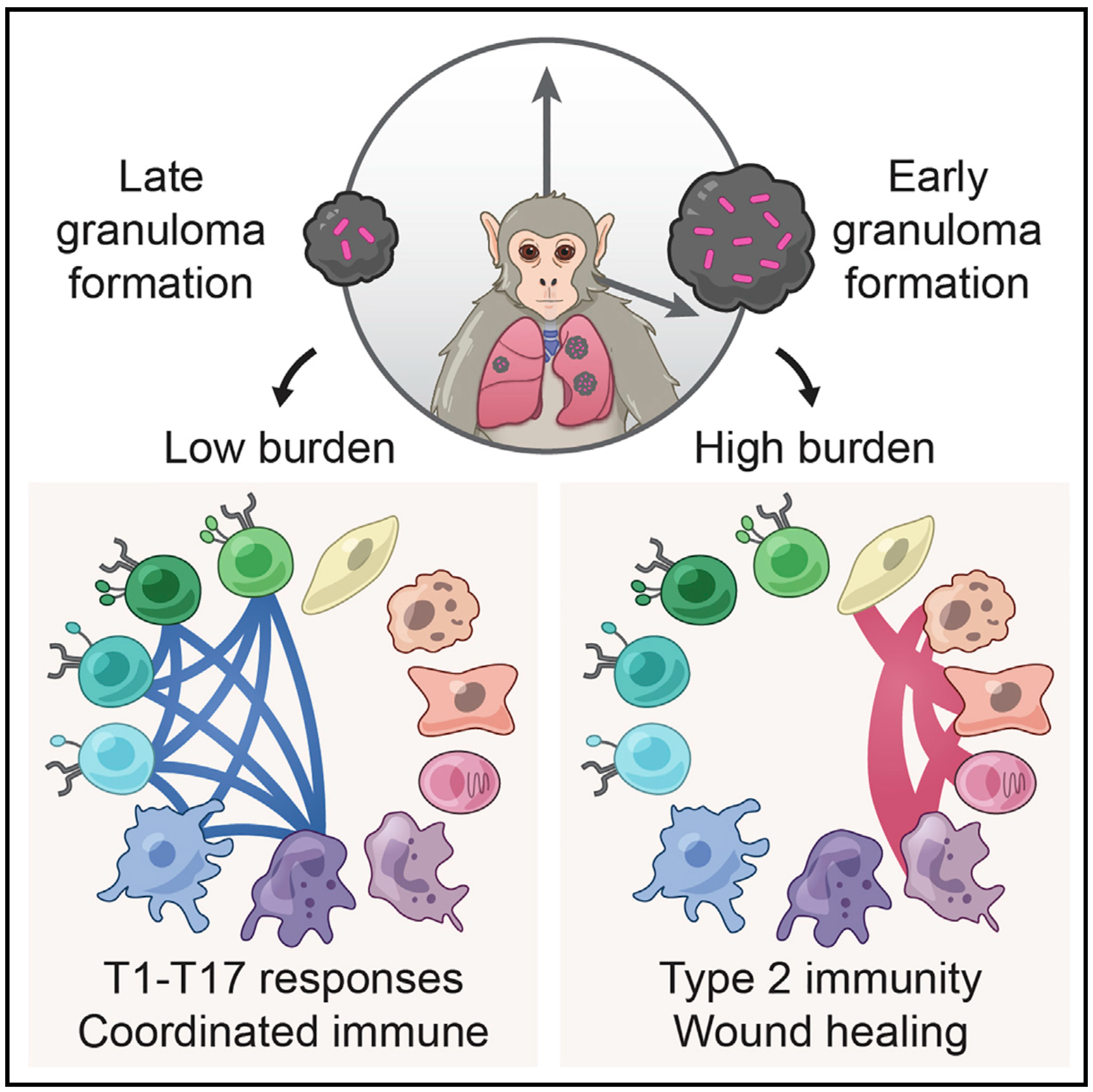
Mycobacterium tuberculosis lung infection results in a complex multicellular structure: the granuloma. In some granulomas, immune activity promotes bacterial clearance, but in others, bacteria persist and grow. We identified correlates of bacterial control in cynomolgus macaque lung granulomas by co-registering longitudinal positron emission tomography and computed tomography imaging, single-cell RNA sequencing, and measures of bacterial clearance. Bacterial persistence occurred in granulomas enriched for mast, endothelial, fibroblast, and plasma cells, signaling amongst themselves via type 2 immunity and wound-healing pathways. Granulomas that drove bacterial control were characterized by cellular ecosystems enriched for type 1-type 17, stem-like, and cytotoxic T cells engaged in pro-inflammatory signaling networks involving diverse cell populations. Granulomas that arose later in infection displayed functional characteristics of restrictive granulomas and were more capable of killing Mtb. Our results define the complex multicellular eco- systems underlying (lack of) granuloma resolution and highlight host immune targets that can be leveraged to develop new vaccine and therapeutic strategies for TB.
Malaria-causing Plasmodium vivax parasites can linger in the human liver for weeks to years and reactivate to cause recurrent blood-stage infection. Although they are an important target for malaria eradication, little is known about the molecular features of replicative and non-replicative intracellular liver-stage parasites and their host cell dependence. Here, we leverage a bioengineered human microliver platform to culture patient-derived P. vivax parasites for transcriptional profiling. Coupling enrichment strategies with bulk and single-cell analyses, we capture both parasite and host transcripts in individual hepatocytes throughout the course of infection. We define host- and state-dependent transcriptional signatures and identify unappreciated populations of replicative and non-replicative parasites that share features with sexual transmissive forms. We find that infection suppresses the transcription of key hepatocyte function genes and elicits an anti-parasite innate immune response. Our work provides a foundation for understanding host-parasite interactions and reveals insights into the biology of P. vivax dormancy and transmission.
Blood samples are frequently collected in human studies of the immune system but poorly represent tissue-resident immunity. Understanding the immunopathogenesis of tissue-restricted diseases, such as chronic hepatitis B, necessitates direct investigation of local immune responses. We developed a workflow that enables frequent, minimally invasive collection of liver fine-needle aspirates in multi-site international studies and centralized single-cell RNA sequencing data generation using the Seq-Well S3 picowell-based technology. All immunological cell types were captured, including liver macrophages, and showed distinct compartmentalization and transcriptional profiles, providing a systematic assessment of the capabilities and limitations of peripheral blood samples when investigating tissue-restricted diseases. The ability to electively sample the liver of chronic viral hepatitis patients and generate high-resolution data will enable multi-site clinical studies to power fundamental and therapeutic discovery.
Mycobacterium tuberculosis (Mtb) bacilli readily aggregate. We previously reported that Mtb aggregates lead to phagocyte death and subsequent efficient replication in the dead infected cells. Here, we examined the transcriptional response of human monocyte derived macrophages to phagocytosis of aggregated Mtb relative to phagocytosis of non-aggregated single or multiple bacilli. Infection with aggregated Mtb led to an early upregulation of pro-inflammatory associated genes and enhanced TNFα signaling via the NFκB pathway. These pathways were significantly more upregulated relative to infection with single or multiple non-aggregated bacilli per cell. Phagocytosis of aggregates led to a decreased phagosome acidification on a per bacillus basis and increased phagocyte cell death, which was not observed when Mtb aggregates were heat killed prior to phagocytosis. Mtb aggregates, observed in a granuloma from a patient, were found surrounding a lesion cavity. These observations suggest that TB aggregation may be a mechanism for pathogenesis. They raise the possibility that aggregated Mtb, if spread from individual to individual, could facilitate increased inflammation, Mtb growth, and macrophage cell death, potentially leading to active disease, cell necrosis, and additional cycles of transmission.
Effective presentation of antigens by HLA class I molecules to CD8+ T cells is required for viral elimination and generation of long-term immunological memory. In this study, we applied a single-cell, multi-omic technology to generate a unified ex vivo characterization of the CD8+ T cell response to SARS-CoV-2 across 4 major HLA class I alleles. We found that HLA genotype conditions key features of epitope specificity, TCR α/β sequence diversity, and the utilization of pre-existing SARS-CoV-2 reactive memory T cell pools. Single-cell transcriptomics revealed functionally diverse T cell phenotypes of SARS-CoV-2-reactive T cells, associated with both disease stage and epitope specificity. Our results show that HLA variations significantly influence the CD8+ T cell repertoire shape and utilization of immune recall upon SARS-CoV-2 infection.
Many patients infected with coronaviruses, such as SARS-CoV-2 and NL63 that use ACE2 receptors to infect cells, exhibit gastrointestinal symptoms and viral proteins are found in the human gastrointestinal tract, yet little is known about the inflammatory and pathological effects of coronavirus infection on the human intestine. Here, we used a human intestine-on-a-chip (Intestine Chip) microfluidic culture device lined by patient organoid-derived intestinal epithelium interfaced with human vascular endothelium to study host cellular and inflammatory responses to infection with NL63 coronavirus. These organoid-derived intestinal epithelial cells dramatically increased their ACE2 protein levels when cultured under flow in the presence of peristalsis-like mechanical deformations in the Intestine Chips compared to when cultured statically as organoids or in Transwell inserts. Infection of the intestinal epithelium with NL63 on-chip led to inflammation of the endothelium as demonstrated by loss of barrier function, increased cytokine production, and recruitment of circulating peripheral blood mononuclear cells (PBMCs). Treatment of NL63 infected chips with the approved protease inhibitor drug, nafamostat, inhibited viral entry and resulted in a reduction in both viral load and cytokine secretion, whereas remdesivir, one of the few drugs approved for COVID19 patients, was not found to be effective and it also was toxic to the endothelium. This model of intestinal infection was also used to test the effects of other drugs that have been proposed for potential repurposing against SARS-CoV-2. Taken together, these data suggest that the human Intestine Chip might be useful as a human preclinical model for studying coronavirus related pathology as well as for testing of potential anti-viral or anti-inflammatory therapeutics.
Prior studies have demonstrated that immunologic dysfunction underpins severe illness in COVID-19 patients, but have lacked an in-depth analysis of the immunologic drivers of death in the most critically ill patients. We performed immunophenotyping of viral antigen-specific and unconventional T cell responses, neutralizing antibodies, and serum proteins in critically ill patients with SARS-CoV-2 infection, using influenza infection, SARS-CoV-2-convalescent health care workers, and healthy adults as controls. We identify mucosal-associated invariant T (MAIT) cell activation as an independent and significant predictor of death in COVID-19 (HR = 5.92, 95% CI = 2.49–14.1). MAIT cell activation correlates with several other mortality-associated immunologic measures including broad activation of CD8+ T cells and non-Vδ2 γδT cells, and elevated levels of cytokines and chemokines, including GM-CSF, CXCL10, CCL2, and IL-6. MAIT cell activation is also a predictor of disease severity in influenza (ECMO/death HR = 4.43, 95% CI = 1.08–18.2). Single-cell RNA-sequencing reveals a shift from focused IFNα-driven signals in COVID-19 ICU patients who survive to broad pro-inflammatory responses in fatal COVID-19 –a feature not observed in severe influenza. We conclude that fatal COVID-19 infection is driven by uncoordinated inflammatory responses that drive a hierarchy of T cell activation, elements of which can serve as prognostic indicators and potential targets for immune intervention.
SARS-CoV-2 infects epithelial cells of the human gastrointestinal (GI) tract and causes related symptoms. HIV infection impairs gut homeostasis and is associated with an increased risk of COVID-19 fatality. To investigate the potential link between these observations, we analyzed single-cell transcriptional profiles and SARS-CoV-2 entry receptor expression across lymphoid and mucosal human tissue from chronically HIV-infected individuals and uninfected controls. Absorptive gut enterocytes displayed the highest coexpression of SARS-CoV-2 receptors ACE2, TMPRSS2, and TMPRSS4, of which ACE2 expression was associated with canonical interferon response and antiviral genes. Chronic treated HIV infection was associated with a clear antiviral response in gut enterocytes and, unexpectedly, with a substantial reduction of ACE2 and TMPRSS2 target cells. Gut tissue from SARS-CoV-2–infected individuals, however, showed abundant SARS-CoV-2 nucleocapsid protein in both the large and small intestine, including an HIV-coinfected individual. Thus, upregulation of antiviral response genes and downregulation of ACE2 and TMPRSS2 in the GI tract of HIV-infected individuals does not prevent SARS-CoV-2 infection in this compartment. The impact of these HIV-associated intestinal mucosal changes on SARS-CoV-2 infection dynamics, disease severity, and vaccine responses remains unclear and requires further investigation.
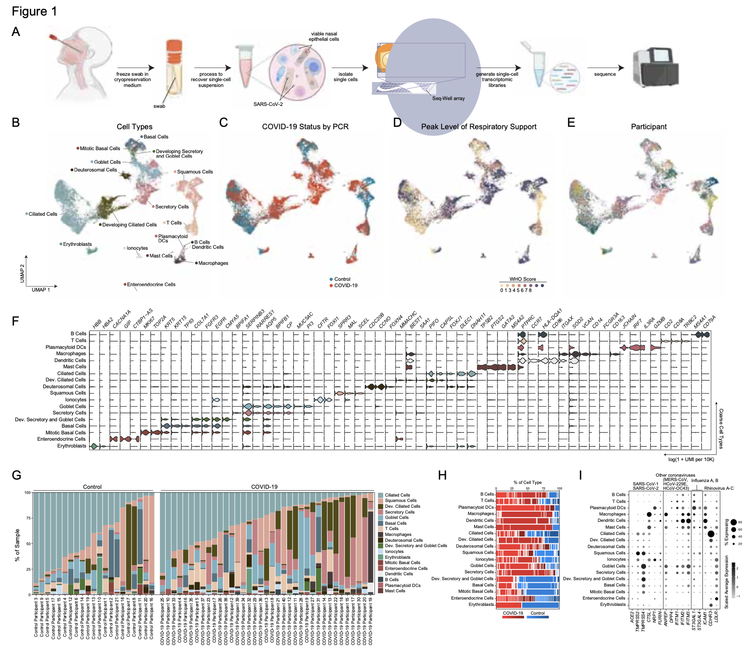
SARS-CoV-2 infection can cause severe respiratory COVID-19. However, many individuals present with isolated upper respiratory symptoms, suggesting potential to constrain viral pathology to the nasopharynx. Which cells SARS-CoV-2 primarily targets and how infection influences the respiratory epithelium remains incompletely understood. We performed scRNA-seq on nasopharyngeal swabs from 58 healthy and COVID-19 participants. During COVID-19, we observe expansion of secretory, loss of ciliated, and epithelial cell repopulation via deuterosomal expansion. In mild/moderate COVID-19, epithelial cells express anti-viral/interferon-responsive genes, while cells in severe COVID-19 have muted anti-viral responses despite equivalent viral loads. SARS-CoV-2 RNA+ host-target cells are highly heterogenous, including developing ciliated, interferon-responsive ciliated, AZGP1high goblet, and KRT13+ “hillock”-like cells, and we identify genes associated with susceptibility, resistance, or infection response. Our study defines protective and detrimental responses to SARS-CoV-2, the direct viral targets of infection, and suggests that failed nasal epithelial anti-viral immunity may underlie and precede severe COVID-19.
Granulomas are complex cellular structures composed predominantly of macrophages and lymphocytes that function to contain and kill invading pathogens. Here, we investigated the single-cell phenotypes associated with antimicrobial responses in human leprosy granulomas by applying single-cell and spatial sequencing to leprosy biopsy specimens. We focused on reversal reactions (RRs), a dynamic process whereby some patients with disseminated lepromatous leprosy (L-lep) transition toward self-limiting tuberculoid leprosy (T-lep), mounting effective antimicrobial responses. We identified a set of genes encoding proteins involved in antimicrobial responses that are differentially expressed in RR versus L-lep lesions and regulated by interferon-γ and interleukin-1β. By integrating the spatial coordinates of the key cell types and antimicrobial gene expression in RR and T-lep lesions, we constructed a map revealing the organized architecture of granulomas depicting compositional and functional layers by which macrophages, T cells, keratinocytes and fibroblasts can each contribute to the antimicrobial response.
The skin lesion erythema migrans (EM) is an initial sign of the Ixodes tick–transmitted Borreliella spirochetal infection known as Lyme disease. T cells and innate immune cells have previously been shown to predominate the EM lesion and promote the reaction. Despite the established importance of B cells and antibodies in preventing infection, the role of B cells in the skin immune response to Borreliella is unknown. Here, we used single-cell RNA-Seq in conjunction with B cell receptor (BCR) sequencing to immunophenotype EM lesions and their associated B cells and BCR repertoires. We found that B cells were more abundant in EM in comparison with autologous uninvolved skin; many were clonally expanded and had circulating relatives. EM-associated B cells upregulated
the expression of MHC class II genes and exhibited preferential IgM isotype usage. A subset also exhibited low levels of somatic hypermutation despite a gene expression profile consistent with memory B cells. Our study demonstrates that single-cell gene expression with paired BCR sequencing can be used to interrogate the sparse B cell populations in human skin and reveals that B cells in the skin infection site in early Lyme disease expressed a phenotype consistent with local antigen presentation and antibody production.
COVID-19, caused by SARS-CoV-2, can result in acute respiratory distress syndrome and multiple-organ failure, but little is known about its pathophysiology. Here, we generated single-cell atlases of 23 lung, 16 kidney, 16 liver and 19 heart COVID-19 autopsy donor tissue samples, and spatial atlases of 14 lung donors. Integrated computational analysis uncovered substantial remodeling in the lung epithelial, immune and stromal compartments, with evidence of multiple paths of failed tissue regeneration, including defective alveolar type 2 differentiation and expansion of fibroblasts and putative TP63+ intrapulmonary basal-like progenitor cells. Viral RNAs were enriched in mononuclear phagocytic and endothelial lung cells which induced specific host programs. Spatial analysis in lung distinguished inflammatory host responses in lung regions with and without viral RNA. Analysis of the other tissue atlases showed transcriptional alterations in multiple cell types in COVID-19 donor heart tissue, and mapped cell types and genes implicated with disease severity based on COVID-19 GWAS. Our foundational dataset elucidates the biological impact of severe SARS-CoV-2 infection across the body, a key step towards new treatments.
Environmental enteropathy (EE) is a subclinical condition of the small intestine that is highly prevalent in low- and middle income countries. It is thought to be a primary cause of most global growth-stunting cases and a key contributing factor to childhood malnutrition and diminished oral vaccine responses. While EE has been shown to be the byproduct of recurrent enteric infection, to date, its full pathophysiology remains unclear. Here, we mapped the cellular and molecular correlates of EE severity by performing high-throughput single-cell RNA-sequencing on 33 small intestinal biopsies from 11 adults with EE from Lusaka, Zambia (8 HIV negative, 3 HIV positive) and 6 adults without EE in Boston, USA. Using the resulting cellular atlas, we scored existing bulk-transcriptomic signatures of reduced villus height and decreased plasma LPS levels in EE, finding that these signatures may be driven by an increased abundance of surface mucosal cells (a gastric-like subset previously implicated in epithelial repair in the gastrointestinal tract). In addition, we identified several cell subsets whose fractional abundances associated with histological determined EE severity, small intestinal region, and HIV infection. Furthermore, by comparing distal duodenal EE samples with those from two U.S. control cohorts, we identified broadly decreased epithelial proliferative signaling, lower fractional abundances of goblet cells, and a T cell subset highly expressing a transcriptional signature of tissue-resident memory cells but with increased pro-inflammatory cytokine expression in EE. Altogether, our work illuminates the epithelial and immune correlates of EE severity and provides new molecular targets for intervention.
Mucosal associated invariant T (MAIT) cells are a class of innate-like T cells that utilize a semi-invariant αβ T cell receptor to recognize small molecule ligands produced by bacteria and fungi. Despite growing evidence that immune cells at mucosal surfaces are often phenotypically and functionally distinct from those in the peripheral circulation, knowledge about the characteristics of MAIT cells at the lung mucosal surface, the site of exposure to respiratory pathogens, is limited. HIV infection has been shown to have a profound effect on the number and function of MAIT cells in the peripheral blood, but its effect on lung mucosal MAIT cells is unknown. We examined the phenotypic, functional, and transcriptomic features of major histocompatibility complex (MHC) class I-related (MR1)-restricted MAIT cells from the peripheral blood and bronchoalveolar compartments of otherwise healthy individuals with latent Mycobacterium tuberculosis (Mtb) infection who were either HIV uninfected or HIV infected. Peripheral blood MAIT cells consistently co-expressed typical MAIT cell surface markers CD161 and CD26 in HIV-negative individuals, while paired bronchoalveolar MAIT cells displayed heterogenous expression of these markers. Bronchoalveolar MAIT cells produced lower levels of pro-inflammatory cytokine IFN-γ and expressed higher levels of co-inhibitory markers PD-1 and TIM-3 than peripheral MAIT cells. HIV infection resulted in decreased frequencies and pro-inflammatory function of peripheral blood MAIT cells, while in the bronchoalveolar compartment MAIT cell frequency was decreased but phenotype and function were not significantly altered. Single-cell transcriptomic analysis demonstrated greater heterogeneity among bronchoalveolar compared to peripheral blood MAIT cells and suggested a distinct subset in the bronchoalveolar compartment. The transcriptional features of this bronchoalveolar subset were associated with MAIT cell tissue repair functions. In summary, we found previously undescribed phenotypic and transcriptional heterogeneity of bronchoalveolar MAIT cells in HIV-negative people. In HIV infection, we found numeric depletion of MAIT cells in both anatomical compartments but preservation of the novel phenotypic and transcriptional features of bronchoalveolar MAIT cells.
Angiotensin-converting enzyme 2 (ACE2) and accessory proteases (TMPRSS2 and CTSL) are needed for severe acute respiratory syndrome coronavirus 2 (SARS-CoV-2) cellular entry, and their expression may shed light on viral tropism and impact across the body. We assessed the cell-type-specific expression of ACE2, TMPRSS2 and CTSL across 107 single-cell RNA-sequencing studies from different tissues. ACE2, TMPRSS2 and CTSL are coexpressed in specific subsets of respiratory epithelial cells in the nasal passages, airways and alveoli, and in cells from other organs associated with coronavirus disease 2019 (COVID-19) transmission or pathology. We performed a meta-analysis of 31 lung single-cell RNA-sequencing studies with 1,320,896 cells from 377 nasal, airway and lung parenchyma samples from 228 individuals. This revealed cell-type-specific associations of age, sex and smoking with expression levels of ACE2, TMPRSS2 and CTSL. Expression of entry factors increased with age and in males, including in airway secretory cells and alveolar type 2 cells. Expression programs shared by ACE2+TMPRSS2+ cells in nasal, lung and gut tissues included genes that may mediate viral entry, key immune functions and epithelial–macrophage cross-talk, such as genes involved in the interleukin-6, interleukin-1, tumor necrosis factor and complement pathways. Cell-type-specific expression patterns may contribute to the pathogenesis of COVID-19, and our work highlights putative molecular pathways for therapeutic intervention.
The SARS-CoV-2 pandemic has caused over 1 million deaths globally, mostly due to acute lung injury and acute respiratory distress syndrome, or direct complications resulting in multiple-organ failures. Little is known about the host tissue immune and cellular responses associated with COVID-19 infection, symptoms, and lethality. To address this, we collected tissues from 11 organs during the clinical autopsy of 17 individuals who succumbed to COVID-19, resulting in a tissue bank of approximately 420 specimens. We generated comprehensive cellular maps capturing COVID-19 biology related to patients’ demise through single-cell and single-nucleus RNA-Seq of lung, kidney, liver and heart tissues, and further contextualized our findings through spatial RNA profiling of distinct lung regions. We developed a computational framework that incorporates removal of ambient RNA and automated cell type annotation to facilitate comparison with other healthy and diseased tissue atlases. In the lung, we uncovered significantly altered transcriptional programs within the epithelial, immune, and stromal compartments and cell intrinsic changes in multiple cell types relative to lung tissue from healthy controls. We observed evidence of: alveolar type 2 (AT2) differentiation replacing depleted alveolar type 1 (AT1) lung epithelial cells, as previously seen in fibrosis; a concomitant increase in myofibroblasts reflective of defective tissue repair; and, putative TP63+ intrapulmonary basal-like progenitor (IPBLP) cells, similar to cells identified in H1N1 influenza, that may serve as an emergency cellular reserve for severely damaged alveoli. Together, these findings suggest the activation and failure of multiple avenues for regeneration of the epithelium in these terminal lungs. SARS-CoV-2 RNA reads were enriched in lung mononuclear phagocytic cells and endothelial cells, and these cells expressed distinct host response transcriptional programs. We corroborated the compositional and transcriptional changes in lung tissue through spatial analysis of RNA profiles in situ and distinguished unique tissue host responses between regions with and without viral RNA, and in COVID-19 donor tissues relative to healthy lung. Finally, we analyzed genetic regions implicated in COVID-19 GWAS with transcriptomic data to implicate specific cell types and genes associated with disease severity. Overall, our COVID-19 cell atlas is a foundational dataset to better understand the biological impact of SARS-CoV-2 infection across the human body and empowers the identification of new therapeutic interventions and prevention strategies.
Infection with SARS-CoV-2, the virus that causes COVID-19, can lead to severe lower respiratory illness including pneumonia and acute respiratory distress syndrome, which can result in profound morbidity and mortality. However, many infected individuals are either asymptomatic or have isolated upper respiratory symptoms, which suggests that the upper airways represent the initial site of viral infection, and that some individuals are able to largely constrain viral pathology to the nasal and oropharyngeal tissues. Which cell types in the human nasopharynx are the primary targets of SARS-CoV-2 infection, and how infection influences the cellular organization of the respiratory epithelium remains incompletely understood. Here, we present nasopharyngeal samples from a cohort of 35 individuals with COVID-19, representing a wide spectrum of disease states from ambulatory to critically ill, as well as 23 healthy and intubated patients without COVID-19. Using standard nasopharyngeal swabs, we collected viable cells and performed single-cell RNA-sequencing (scRNA-seq), simultaneously profiling both host and viral RNA. We find that following infection with SARS-CoV-2, the upper respiratory epithelium undergoes massive reorganization: secretory cells diversify and expand, and mature epithelial cells are preferentially lost. Further, we observe evidence for deuterosomal cell and immature ciliated cell expansion, potentially representing active repopulation of lost ciliated cells through coupled secretory cell differentiation. Epithelial cells from participants with mild/moderate COVID-19 show extensive induction of genes associated with anti-viral and type I interferon responses. In contrast, cells from participants with severe lower respiratory symptoms appear globally muted in their anti-viral capacity, despite substantially higher local inflammatory myeloid populations and equivalent nasal viral loads. This suggests an essential role for intrinsic, local epithelial immunity in curbing and constraining viral-induced pathology. Using a custom computational pipeline, we characterized cell-associated SARS-CoV-2 RNA and identified rare cells with RNA intermediates strongly suggestive of active replication. Both within and across individuals, we find remarkable diversity and heterogeneity among SARS-CoV-2 RNA+ host cells, including developing/immature and interferon-responsive ciliated cells, KRT13+ “hillock”-like cells, and unique subsets of secretory, goblet, and squamous cells. Finally, SARS-CoV-2 RNA+ cells, as compared to uninfected bystanders, are enriched for genes involved in susceptibility (e.g., CTSL, TMPRSS2) or response (e.g., MX1, IFITM3, EIF2AK2) to infection. Together, this work defines both protective and detrimental host responses to SARS-CoV-2, determines the direct viral targets of infection, and suggests that failed anti-viral epithelial immunity in the nasal mucosa may underlie the progression to severe COVID-19.
In late 2019 and through 2020, the COVID-19 pandemic swept the world, presenting both scientific and medical challenges associated with understanding and treating a previously unknown disease. To help address the need for great understanding of COVID-19, the scientific community mobilized and banded together rapidly to characterize SARS-CoV-2 infection, pathogenesis and its distinct disease trajectories. The urgency of COVID-19 provided a pressing use-case for leveraging relatively new tools, technologies, and nascent collaborative networks. Single-cell biology is one such example that has emerged over the last decade as a powerful approach that provides unprecedented resolution to the cellular and molecular underpinnings of biological processes. Early foundational work within the single-cell community, including the Human Cell Atlas, utilized published and unpublished data to characterize the putative target cells of SARS-CoV-2 sampled from diverse organs based on expression of the viral receptor ACE2 and associated entry factors TMPRSS2 and CTSL (Muus et al., 2020; Sungnak et al., 2020; Ziegler et al., 2020). This initial characterization of reference data provided an important foundation for framing infection and pathology in the airway as well as other organs. However, initial community analysis was limited to samples derived from uninfected donors and other previously-sampled disease indications. This report provides an overview of a single-cell data resource derived from samples from COVID-19 patients along with initial observations and guidance on data reuse and exploration.
Granulomas are complex cellular structures comprised predominantly of macrophages and lymphocytes that function to contain and kill invading pathogens. Here, we investigated single cell phenotypes associated with antimicrobial responses in human leprosy granulomas by applying single cell and spatial sequencing to leprosy biopsy specimens. We focused on reversal reactions (RR), a dynamic process in which some patients with disseminated lepromatous leprosy (L-lep) transition towards self-limiting tuberculoid leprosy (T-lep), mounting effective antimicrobial responses. We identified a set of genes encoding proteins involved in antimicrobial responses that are differentially expressed in RR versus L-lep lesions, and regulated by IFN-γ and IL-1β. By integrating the spatial coordinates of the key cell types and antimicrobial gene expression in RR and T-lep lesions, we constructed a map revealing the organized architecture of granulomas depicting compositional and functional layers by which macrophages, T cells, keratinocytes and fibroblasts contribute to the antimicrobial response.
Understanding the earliest immune responses following HIV infection is critical to inform future vaccines and therapeutics. Here, we review recent prospective human studies in at-risk populations that have provided insight into immune responses during acute infection, including additional relevant data from non-human primate (NHP) studies. We discuss the timing, nature, and function of the diverse immune responses induced, the onset of immune dysfunction, and the effects of early anti-retroviral therapy administration. Treatment at onset of viremia mitigates peripheral T and B cell dysfunction, limits seroconversion, and enhances cellular antiviral immunity despite persistence of infection in lymphoid tissues. We highlight pertinent areas for future investigation, and how application of high-throughput technologies, alongside targeted NHP studies, may elucidate immune response features to target in novel preventions and cures.
Ebola virus (EBOV) causes epidemics with high mortality yet remains understudied due to the challenge of experimentation in high-containment and outbreak settings. Here, we used single-cell transcriptomics and CyTOF-based single-cell protein quantification to characterize peripheral immune cells during EBOV infection in rhesus monkeys. We obtained 100,000 transcriptomes and 15,000,000 protein profiles, finding that immature, proliferative monocyte-lineage cells with reduced antigen-presentation capacity replace conventional monocyte subsets, while lymphocytes upregulate apoptosis genes and decline in abundance. By quantifying intracellular viral RNA, we identify molecular determinants of tropism among circulating immune cells and examine temporal dynamics in viral and host gene expression. Within infected cells, EBOV downregulates STAT1 mRNA and interferon signaling, and it upregulates putative pro-viral genes (e.g., DYNLL1 and HSPA5), nominating pathways the virus manipulates for its replication. This study sheds light on EBOV tropism, replication dynamics, and elicited immune response and provides a framework for characterizing host-virus interactions under maximum containment.
In humans and nonhuman primates, Mycobacterium tuberculosis lung infection yields a complex multicellular structure: the tuberculosis granuloma. All granulomas are not equivalent, however, even within the same host: in some, local immune activity promotes bacterial clearance, while in others, it allows persistence or outgrowth. Here, we used single-cell RNA-sequencing to define holistically cellular responses associated with control in cynomolgus macaques. Granulomas that facilitated bacterial killing contained significantly higher proportions of CD4+ and CD8+ T cells expressing hybrid Type1-Type17 immune responses or stem-like features and CD8-enriched T cells with specific cytotoxic functions; failure to control correlated with mast cell, plasma cell and fibroblast abundance. Co-registering these data with serial PET-CT imaging suggests that a degree of early immune control can be achieved through cytotoxic activity, but that more robust restriction only arises after the priming of specific adaptive immune responses, defining new targets for vaccination and treatment.
Innate lymphoid cells (ILCs) are important for response to infection and for immune development in early life. HIV infection in adults depletes circulating ILCs, but the impact on children infected from birth remains unknown. We study vertically HIV-infected children from birth to adulthood and find severe and persistent depletion of all circulating ILCs that, unlike CD4+ T cells, are not restored by long-term antiretroviral therapy unless initiated at birth. Remaining ILCs upregulate genes associated with cellular activation and metabolic perturbation. Unlike HIV-infected adults, ILCs are also profoundly depleted in tonsils of vertically infected children. Transcriptional profiling of remaining ILCs reveals ongoing cell-type-specific activity despite anti-retroviral therapy. Collectively, these data suggest an important and ongoing role for ILCs in lymphoid tissue of HIV-infected children from birth, where persistent depletion and sustained transcriptional activity are likely to have long-term immune consequences that merit further investigation.
Our nasal epithelial COVID-19 dataset, along with COVID-19 datasets from other genomics groups, can now be found at covid19cellatlas.org. This work was sponsored by the Chan-Zuckerberg Initiative.
B cell receptors (BCRs) display a combination of variable (V)-gene-encoded complementarity determining regions (CDRs) and adaptive/hypervariable CDR3 loops to engage antigens. It has long been proposed that the former tune for recognition of pathogens or groups of pathogens. To experimentally evaluate this within the human antibody repertoire, we perform immune challenges in transgenic mice that bear diverse human CDR3 and light chains but are constrained to different human VH–genes. We find that, of six commonly deployed VHsequences, only those CDRs encoded by IGHV1-2∗02 enable polyclonal antibody responses against bacterial lipopolysaccharide (LPS) when introduced to the bloodstream. The LPS is from diverse strains of gram-negative bacteria, and the VH-gene-dependent responses are directed against the non-variable and universal saccrolipid substructure of this antigen. This reveals a broad-spectrum anti-LPS response in which germline-encoded CDRs naturally hardwire the human antibody repertoire for recognition of a conserved microbial target.
Ebola virus (EBOV) causes epidemics with high case fatality rates, yet remains understudied due to the challenge of experimentation in high-containment and outbreak settings. To better understand EBOV infection in vivo, we used single-cell transcriptomics and CyTOF-based single-cell protein quantification to characterize peripheral immune cell activity during EBOV infection in rhesus monkeys. We obtained 100,000 transcriptomes and 15,000,000 protein profiles, providing insight into pathogenesis. We find that immature, proliferative monocyte-lineage cells with reduced antigen presentation capacity replace conventional circulating monocyte subsets within days of infection, while lymphocytes upregulate apoptosis genes and decline in abundance. By quantifying viral RNA abundance in individual cells, we identify molecular determinants of tropism and examine temporal dynamics in viral and host gene expression. Within infected cells, we observe that EBOV down-regulates STAT1 mRNA and interferon signaling, and up-regulates putative pro-viral genes (e.g., DYNLL1 and HSPA5), nominating cellular pathways the virus manipulates for its replication. Overall, this study sheds light on EBOV tropism, replication dynamics, and elicited immune response, and provides a framework for characterizing interactions between hosts and emerging viruses in a maximum containment setting.
We investigated SARS-CoV-2 potential tropism by surveying expression of viral entry-associated genes in single-cell RNA-sequencing data from multiple tissues from healthy human donors. We co-detected these transcripts in specific respiratory, corneal and intestinal epithelial cells, potentially explaining the high efficiency of SARS-CoV-2 transmission. These genes are co-expressed in nasal epithelial cells with genes involved in innate immunity, highlighting the cells’ potential role in initial viral infection, spread and clearance. The study offers a useful resource for further lines of inquiry with valuable clinical samples from COVID-19 patients and we provide our data in a comprehensive, open and user-friendly fashion at www.covid19cellatlas.org.
There is pressing urgency to understand the pathogenesis of the severe acute respiratory syndrome coronavirus clade 2 (SARS-CoV-2) which causes the disease COVID-19. SARS-CoV- 2 spike (S)-protein binds ACE2, and in concert with host proteases, principally TMPRSS2, promotes cellular entry. The cell subsets targeted by SARS-CoV-2 in host tissues, and the factors that regulate ACE2 expression, remain unknown. Here, we leverage human, non-human primate, and mouse single-cell RNA-sequencing (scRNA-seq) datasets across health and disease to uncover putative targets of SARS-CoV-2 amongst tissue-resident cell subsets. We identify ACE2 and TMPRSS2 co-expressing cells within lung type II pneumocytes, ileal absorptive enterocytes, and nasal goblet secretory cells. Strikingly, we discover that ACE2 is a human interferon- stimulated gene (ISG) in vitro using airway epithelial cells, and extend our findings to in vivo viral infections. Our data suggest that SARS-CoV-2 could exploit species-specific interferon-driven upregulation of ACE2, a tissue-protective mediator during lung injury, to enhance infection.
The COVID-19 pandemic, caused by the novel coronavirus SARS-CoV-2, creates an urgent need for identifying molecular mechanisms that mediate viral entry, propagation, and tissue pathology. Cell membrane bound angiotensin-converting enzyme 2 (ACE2) and associated proteases, transmembrane protease serine 2 (TMPRSS2) and Cathepsin L (CTSL), were previously identified as mediators of SARS-CoV2 cellular entry. Here, we assess the cell type-specific RNA expression of ACE2, TMPRSS2, and CTSL through an integrated analysis of 107 single-cell and single-nucleus RNA-Seq studies, including 22 lung and airways datasets (16 unpublished), and 85 datasets from other diverse organs. Joint expression of ACE2 and the accessory proteases identifies specific subsets of respiratory epithelial cells as putative targets of viral infection in the nasal passages, airways, and alveoli. Cells that co-express ACE2 and proteases are also identified in cells from other organs, some of which have been associated with COVID-19 transmission or pathology, including gut enterocytes, corneal epithelial cells, cardiomyocytes, heart pericytes, olfactory sustentacular cells, and renal epithelial cells. Performing the first meta- analyses of scRNA-seq studies, we analyzed 1,176,683 cells from 282 nasal, airway, and lung parenchyma samples from 164 donors spanning fetal, childhood, adult, and elderly age groups, associate increased levels of ACE2, TMPRSS2, and CTSL in specific cell types with increasing age, male gender, and smoking, all of which are epidemiologically linked to COVID-19 susceptibility and outcomes. Notably, there was a particularly low expression of ACE2 in the few young pediatric samples in the analysis. Further analysis reveals a gene expression program shared by ACE2+TMPRSS2+ cells in nasal, lung and gut tissues, including genes that may mediate viral entry, subtend key immune functions, and mediate epithelial-macrophage cross- talk. Amongst these are IL6, its receptor and co-receptor, IL1R, TNF response pathways, and complement genes. Cell type specificity in the lung and airways and smoking effects were conserved in mice. Our analyses suggest that differences in the cell type-specific expression of mediators of SARS-CoV-2 viral entry may be responsible for aspects of COVID-19 epidemiology and clinical course, and point to putative molecular pathways involved in disease susceptibility and pathogenesis.
Influenza virus infections are major causes of morbidity and mortality. Research using cultured cells, bulk tissue, and animal models cannot fully capture human disease dynamics. Many aspects of virus-host interactions in a natural setting remain unclear, including the specific cell types that are infected and how they and neighboring bystander cells contribute to the overall antiviral response. To address these questions, we performed single-cell RNA sequencing (scRNA-Seq) on cells from freshly collected nasal washes from healthy human donors and donors diagnosed with acute influenza during the 2017-18 season. We describe a previously uncharacterized goblet cell population, specific to infected individuals, with high expression of MHC class II genes. Furthermore, leveraging scRNA-Seq reads, we obtained deep viral genome coverage and developed a model to rigorously identify infected cells that detected influenza infection in all epithelial cell types and even some immune cells. Our data revealed that each donor was infected by a unique influenza variant and that each variant was separated by at least one unique non-synonymous difference. Our results demonstrate the power of massively-parallel scRNA-Seq to study viral variation, as well as host and viral transcriptional activity during human infection.
Cellular immunity is critical for controlling intracellular pathogens, but individual cellular dynamics and cell–cell cooperativity in evolving human immune responses remain poorly understood. Single-cell RNA-sequencing (scRNA-seq) represents a powerful tool for dissecting complex multicellular behaviors in health and disease and nominating testable therapeutic targets. Its application to longitudinal samples could afford an opportunity to uncover cellular factors associated with the evolution of disease progression without potentially confounding inter-individual variability. Here, we present an experimental and computational methodology that uses scRNA-seq to characterize dynamic cellular programs and their molecular drivers, and apply it to HIV infection. By performing scRNA-seq on peripheral blood mononuclear cells from four untreated individuals before and longitudinally during acute infection, we were powered within each to discover gene response modules that vary by time and cell subset. Beyond previously unappreciated individual- and cell-type-specific interferon-stimulated gene upregulation, we describe temporally aligned gene expression responses obscured in bulk analyses, including those involved in proinflammatory T cell differentiation, prolonged monocyte major histocompatibility complex II upregulation and persistent natural killer (NK) cell cytolytic killing. We further identify response features arising in the first weeks of infection, for example proliferating natural killer cells, which potentially may associate with future viral control. Overall, our approach provides a unified framework for characterizing multiple dynamic cellular responses and their coordination.
There is pressing urgency to better understand the pathogenesis of the severe acute respiratory syndrome (SARS) coronavirus (CoV) clade SARS-CoV-2, which causes the disease known as COVID-19. SARS-CoV-2, like SARS-CoV, utilizes ACE2 to bind host cells. While initial SARS- CoV-2 cell entry and infection depend on ACE2 in concert with the protease TMPRSS2 for spike (S) protein activation, the specific cell subsets targeted by SARS-CoV-2 in host tissues, and the factors that regulate ACE2 expression, remain unknown. Here, we leverage human and non- human primate (NHP) single-cell RNA-sequencing (scRNA-seq) datasets to uncover the tissue- resident cell subsets that may serve as the cellular targets of SARS-CoV-2. We identify ACE2 and TMPRSS2 co-expressing cells within type II pneumocytes in NHP lung, absorptive enterocytes in human and NHP terminal ileum, and human nasal goblet secretory cells. Strikingly, we discover, and extensively corroborate using publicly available data sets, that ACE2 is an interferon-stimulated gene (ISG) in human epithelial cells. We further validate this finding in primary upper airway human respiratory epithelial cells. Thus, SARS-CoV-2 may exploit IFN- driven upregulation of ACE2, a key tissue-protective mediator during lung injury, to enhance infection.
Overview
In light of the global effort to better understand the new SARS-CoV-2 virus, we and other researchers from the HCA Lung Biological Network and beyond have begun an initiative to investigate datasets from relevant tissues profiled as part of other ongoing studies. These studies represent, for example, large efforts to characterize HIV, Mtb, and influenza infection and allergy in primary human and non-human primate samples. This page serves as a guide to viewing our data interactively on and downloading datasets from our single-cell portal, the Alexandria Project. Alternatively, bulk downloading of our data is available here. For more on research initiatives in COVID-19 being undertaken by the HCA, and the HCA Lung Biological Network in particular, please visit the HCA website here.
We investigated two genes whose protein products are central to the cellular entry of SARS-CoV-2: ACE2 and TMPRSS2. Consistent with previous studies, we found that the gene encoding ACE2, the SARS-CoV-2 entry receptor, is expressed on a subset of lung epithelial cells, type 2 pneumocytes, and a subset of ileal epithelial cells, absorptive enterocytes, across several datasets. The protease TMPRSS2 primes the spike protein of SARS-CoV-2 and is also important for viral entry. Because of this, we identified cells which co-express ACE2 and TMPRSS2 in our datasets, and investigated additional genes enriched within ACE2 and TMPRSS2 co-expressing cells. As we believe this data may prove useful to other researchers investigating similar questions, we have made our datasets public through the interactive Alexandria Project. Here, you can view our annotations of these datasets and investigate which other genes are highly expressed in these cell subsets of interest.
NB None of the datasets presented here were designed to answer specific questions about COVID-19. Additional studies will be required across larger, appropriately structured cohorts. Further, we provide a note of caution when interpreting scRNA-seq data for low abundance transcripts like ACE2 and TMPRSS2 as detection inefficiencies and/or sequencing depth may result in an underestimation of the actual frequencies of ACE2+ or ACE2+/TMPRSS2+ cells in tissues. Moreover, the protein levels of each may differ from their mRNA abundances. We present each data set separately, as each study differed by method of tissue processing and collection protocols, each of which can influence the frequency of recovered cell subsets.
Analysis
Our pre-print “SARS-CoV-2 receptor ACE2 is an interferon-stimulated gene in human airway epithelial cells and is enriched in specific cell subsets across tissues” can be found here, and the abstract is reproduced below.
There is pressing urgency to better understand the pathogenesis of the severe acute respiratory syndrome (SARS) coronavirus (CoV) clade SARS-CoV-2. SARS-CoV-2, like SARS-CoV, utilizes ACE2 to bind host cells. While initial SARS-CoV-2 cell entry and infection depend on ACE2 in concert with the protease TMPRSS2 for spike (S) protein activation, the specific cell subsets targeted by SARS-CoV-2 in host tissues, and the factors that regulate ACE2 expression, remain unknown. Here, we leverage human and non-human primate (NHP) single-cell RNA-sequencing (scRNA-seq) datasets to uncover the cell subsets that may serve as cellular targets of SARS-CoV-2. We identify ACE2/TMPRSS2 co-expressing cells within type II pneumocytes, absorptive enterocytes, and nasal goblet secretory cells. Strikingly, we discover that ACE2 is an interferon-stimulated gene (ISG) in human barrier tissue epithelial cells. Thus, SARS-CoV-2 may exploit IFN-driven upregulation of ACE2, a key tissue-protective mediator during lung injury, to enhance infection.
Datasets
Atlas of ACE2 expression in healthy non-human primate lung and ileum
In this study, we collected cells from various tissues in healthy and SHIV-infected non-human primates using Seq-Well v1. Here we highlight the lung and ileum and show that ACE2 and TMPRSS2 are co-expressed most frequently in type II pneumocytes in the lung and absorptive enterocytes in the ileum.
To visualize these cells in the Alexandria Project, visit this study: Atlas of healthy non-human primate lung and ileum ACE2+ cells
This project contains two UMAP visualizations (one for lung cells and one for ileum cells): toggle between them by clicking on the ‘Explore’ tab, select ‘View Options’ in the top right hand corner, and switch between lung and ileum under the ‘Load Cluster’ dropdown.
For each of the lung and ileum cell UMAPs, we provide subsets of these visualizations which contain only the cell types enriched for double positive cells. By selecting the ‘Load cluster’ options called ‘Lung Epithelial Cells’ or ‘Ileum Absorptive Enterocytes’, you will be able to view gene expression differences between double positive cells and other cells within those cell types. In the lung epithelial cells visualization, the following additional annotations are available under the ‘Select Annotation’ dropdown:
- ACE2+ – cells expressing ACE2
- TMPRSS2+ – cells expressing TMPRSS2
- ACE2_TMPRSS2_double_positive – cells expressing both genes
- celltype_double_positive – the cell type column and double positive column combined so genes that are differentially expressed between only one cell type may be viewed
- celltype_ACE2+ – same as above for ACE2 expression
After selecting one of these annotations, you can then search for a gene of interest in the ‘Search Genes’ box in the left corner to view the expression of that gene as a violin plot split by the annotation you select. For example, to reproduce Figure 1D in the manuscript, you would select annotation “ACE2_TMPRSS2_double_positive” and search for gene ‘IFNGR2’ in the ‘Search Genes’ box.
The same options are available for the ileal absorptive enterocytes visualization except that the cell type is combined with the double positive/ACE2 column since this subset only includes one cell type.
Full expression matrices (and therefore gene expression values visible when using the ‘Search Genes’ box) are available only for epithelial cell types as this data is derived from a pre-publication study.
Atlas of ACE2 and TMPRSS2 in human ileum
In this study, samples from human ileum were collected, processed, and run on 10x 3′ v2 show ACE2 and TMPRSS2 co-expression in absorptive enterocytes.
To visualize these cells in the Alexandria Portal, visit this study: ACE2 and TMPRSS2 most enriched within GSTA1+MGST3+ absorptive enterocytes in context of non-inflamed terminal ileum
Two tSNE visualizations are available for this study, one of all cells in the ileum samples “non-inflammed-tsne” and one of just the epithelial cells in the samples “non-inflammed-epth-tsne”. To toggle between these, select the “Explore” tab under the study title, then select ‘View options’ from the upper right corner of the plot and choose the visualization of interest in the ‘Load cluster’ dropdown.
Full expression matrices (and therefore gene expression values visible when using the ‘search genes’ box) are available only for the epithelial cell types as this data is derived from a pre-publication study.
Atlas of ACE2 and TMPRSS2 expression in human HIV- and TB-infected lung
In this study, samples from lung surgeries were run with Seq-Well S^3 and contain a variety of immune and epithelial cell types. We found the majority of ACE2 and TPRSS2 double positive cells in type II pneumocyte cells.
To view these cells in the Alexandria Project, visit this study: Human lung HIV-TB co-infection ACE2+ cells
To visualize the double positive cells in these samples, click the ‘Explore’ tab, then select ‘View Options’ in the top right corner of the plot and choose ‘ACE2_TMPRSS2_double_positive’ under the ‘Select Annotation’ dropdown menu.
Full expression matrices (and therefore gene expression values visible when using the ‘search genes’ box) are available only for epithelial cell types as this data is derived from a pre-publication study.
A subset of ACE2+ secretory cells in human nasal mucosa
In this study, we find a subset of secretory cells which co-express ACE2 and TMPRSS2.
To visualize these cells on the Alexandria Project, visit this study: Allergic inflammatory memory in human respiratory epithelial progenitor cells
To view the tSNE of epithelial cells, select the ‘Explore’ tab, select ‘View Options’ in the right hand corner of the plot and choose ‘Epithelial cells’ in the ‘Load Cluster’ dropdown. To view the cell type annotations in the first panel of the above plot choose ‘subset’ in the ‘Select Annotation’ dropdown menu. To view the ACE2/TMPRSS2 double positive cells, select ‘ACE2_TMPRSS2’ from the ‘Select annotation dropdown menu, and to view the cluster subsets from the third panel of this plot, select ‘res_0_8’ from the ‘Select Annotation Dropdown Menu’.
ACE2 and TPRSS2 co-expressing cells found in non-human primate granulomas and adjacent uninvolved lung tissue
In this study, we collected lung tissue from non-human primates infected with mTB. These tissues come from both mTB granulomas and adjacent uninvolved lung in the same monkey.
These cells, profiled using Seq-Well S^3, can be investigated interactively in the Alexandria Project: Epithelial cells in NHP TB granuloma and uninvolved lung
To view the data colored by granuloma and uninvolved lung choose the “Granuloma” annotation accessible by clicking “View Options” in the top right-hand corner and selecting from the “Select Annotation” dropdown menu.
Full expression matrices (and therefore gene expression values visible when using the ‘Search Genes’ box) are available only for relevant cell types as this data is derived from a pre-publication study.
Comparison of ACE2 and TMPRSS2 expression in human duodenal and ileal tissue and organoid-derived epithelial cells
In this study, samples from adult human duodenum and ileum were collected and split for primary tissue single-cell RNA-seq and organoid culture under several conditions and profiled with Seq-Well S^3. Organoids were cultured and passaged every 6-8 days in Matrigel domes with established media conditions meant to recapitulate the broad diversity of in vivo epithelial cell types (Fujii, M., et al., Cell Stem Cell. 2018). Organoid culture media contained recombinant Noggin, Rspondin-3, FGF2, IGF1, afamin-Wnt3A, in addition to Gastrin and TGF-b inhibitor A83-01 with and without recombinant EGF (E/NR3+F2I1Gi+Af-W3+A83). Cells co-expressing ACE2 and TMPRSS2 were identified principally within enterocyte clusters of both tissues and organoids.
Visualize these samples interactively and read more about this study on the Alexandria Project: Comparison of ACE2 and TMPRSS2 expression in human duodenal and ileal tissue and organoid-derived epithelial cells
This project contains two UMAP visualizations (one for organoid cells and one for primary tissue cells): toggle between them by clicking on the ‘Explore’ tab, select ‘View Options’ in the top right hand corner, and switch between tissue and organoid under the ‘Load Cluster’ dropdown.
To visualize cells which co-express ACE2 and TMPRSS2, select the ‘ACE2_TMPRSS2’ option under the ‘Select Annotation’ dropdown. You can then search for a gene of interest in the ‘Search Genes’ box in the left corner to view the expression of that gene as a violin plot split by the annotation you select.
Full expression matrices (and therefore gene expression values visible when using the ‘Search Genes’ box) are available only for enterocyte cell types as this data is derived from a pre-publication study. Expression of ACE2 and TMPRSS2 are available for all cells.
Interferon regulation of ACE2 in human and murine basal cells
Analysis of these datasets and others lead to the hypothesis that expression of the ACE2 receptor may be upregulated by interferon. To further interrogate this hypothesis, we cultured basal cells from two primary human donors, one human basal cell line, and one mouse trachea and stimulated them with IL4, IL17a, IFNgamma, IFNαlpha, IFNbeta for 12 hours overnight. We performed bulk RNA sequencing and differential expression to show a dose dependent upregulation of canonical ISGs (interferon signaling genes). Specifically, we see that ACE2 is most significantly unregulated following IFN alpha stimulation in primary human basal cells, diminished in the BEAS-2B cell line and not seen in mouse cells.
To visualize these samples, the gene expression data may be viewed interactively in the Alexandria Project: Interferon regulation of ACE2 in human and murine basal cells
Under the ‘Explore’ tab, use the ‘Search genes’ field in the top left corner to visualize log-normalized gene expression. To visualize expression in each sample, select ‘View Options’ in the top left corner of the plot and choose the sample of interest under ‘Load cluster’. The ‘Stim_Dose’ annotation refers to the dose of each stimulation condition applied to that sample.
Data for all samples in this study can be dowloaded under the ‘Download’ tab.
Murine nasal mucosa after intranasal interferon exposure
We test the impact of IFNalpha stimulation in vivo by treating two mice intranasally with 200 ng of IFNalpha and two with saline. After 12 hours, the nasal mucosa of the respiratory and olfactory epithelia and underlying lamina propria were isolated and prepared for sequencing with Seq-Well S^3.
These cells may be viewed interactively in the Alexandria Project: Murine nasal mucosa after intranasal interferon exposure
Data for all samples in this study can be dowloaded under the ‘Download’ tab.
Alexandria Project Details
Single Cell Portal Documentation
We thank the Broad Institute Single Cell Portal team for creating the platform that allows Alexandria to exist and for their working tirelessly to help us share our datasets for others to access.
Memories of previous immune events enable barrier tissues to rapidly recall distinct environmental exposures. To effectively inform future responses, these past experiences can be stored in cell types that are long-term residents or essential constituents of tissues. There is an emerging understanding that, in addition to antigen-specific immune cells, diverse haematopoietic, stromal, parenchymal and neuronal cell types can store inflammatory memory. Here, we explore the impact of previous immune activity on various cell lineages with the goal of presenting a unified view of inflammatory memory to environmental exposures (such as allergens, antigens, noxious agents and microorganisms) at barrier tissues. We propose that inflammatory memory is distributed across diverse cell types and stored through shifts in cell states, and we provide a framework to guide future experiments. This distribution and storage may promote adaptation or maladaptation in homeostatic, maintenance and disease settings — especially if the distribution of memory favours cellular cooperation during storage or recall.
The induction of broadly neutralizing antibodies (bnAbs) is highly desired for an effective vaccine against HIV-1. Typically, bnAbs develop in patients with high viremia, but they can also evolve in some untreated HIV-1 controllers with low viral loads. Here, we identify a subgroup of neutralizer-controllers characterized by myeloid DCs (mDCs) with a distinct inflammatory signature and a superior ability to prime T follicular helper (Tfh)-like cells in an STAT4-dependent fashion. This distinct immune profile is associated with a higher frequency of Tfh-like cells in peripheral blood (pTfh) and an enrichment for Tfh-defining genes in circulating CD4+ T cells. Correspondingly, monocytes from this neutralizer controller subgroup upregulate genes encoding for chemotaxis and inflammation, and they secrete high levels of IL-12 in response to TLR stimulation. Our results suggest the existence of multi-compartment immune networks between mDCs, Tfh, and monocytes that may facilitate the development of bnAbs in a subgroup of HIV-1 controllers.
Toxoplasma gondii chronically infects a quarter of the world’s population, and its recrudescence can cause life-threatening disease in immunocompromised individuals and recurrent ocular lesions in the immunocompetent. Acute-stage tachyzoites differentiate into chronic-stage bradyzoites, which form intracellular cysts resistant to immune clearance and existing therapies. The molecular basis of this differentiation is unknown, despite being efficiently triggered by stresses in culture. Through Cas9-mediated screening and single-cell profiling, we identify a Myb-like transcription factor (BFD1) necessary for differentiation in cell culture and in mice. BFD1 accumulates during stress and its synthetic expression is sufficient to drive differentiation. Consistent with its function as a transcription factor, BFD1 binds the promoters of many stage-specific genes and represents a counterpoint to the ApiAP2 factors that dominate our current view of parasite gene regulation. BFD1 provides a genetic switch to study and control Toxoplasma differentiation and will inform prevention and treatment of chronic infections.
Mycobacterium tuberculosis (Mtb) is the leading cause of death from infection worldwide. The only available vaccine, BCG (Bacillus Calmette–Guérin), is given intradermally and has variable efficacy against pulmonary tuberculosis, the major cause of mortality and disease transmission. Here we show that intravenous administration of BCG profoundly alters the protective outcome of Mtb challenge in non-human primates (Macaca mulatta). Compared with intradermal or aerosol delivery, intravenous immunization induced substantially more antigen-responsive CD4 and CD8 T cell responses in blood, spleen, bronchoalveolar lavage and lung lymph nodes. Moreover, intravenous immunization induced a high frequency of antigen-responsive T cells across all lung parenchymal tissues. Six months after BCG vaccination, macaques were challenged with virulent Mtb. Notably, nine out of ten macaques that received intravenous BCG vaccination were highly protected, with six macaques showing no detectable levels of infection, as determined by positron emission tomography–computed tomography imaging, mycobacterial growth, pathology and granuloma formation. The finding that intravenous BCG prevents or substantially limits Mtb infection in highly susceptible rhesus macaques has important implications for vaccine delivery and clinical development, and provides a model for defining immune correlates and mechanisms of vaccine-elicited protection against tuberculosis.
Antibody paratopes are formed by hypervariable complementarity-determining regions (CDRH3s) and variable gene-encoded CDRs. The latter show biased usage in human broadly neutralizing antibodies (bnAbs) against both HIV and influenza virus, suggesting the existence of gene-endowed targeting solutions that may be amenable to pathway amplification. To test this, we generated transgenic mice with human CDRH3 diversity but simultaneously constrained to individual user-defined human immunoglobulin variable heavy-chain (VH) genes, including IGHV1-69, which shows biased usage in human bnAbs targeting the hemagglutinin stalk of group 1 influenza A viruses. Sequential immunization with a stalk-only hemagglutinin nanoparticle elicited group 1 bnAbs, but only in IGHV1-69 mice. This VH-endowed response required minimal affinity maturation, was elicited alongside pre-existing influenza immunity, and when IGHV1-69 B cells were diluted to match the frequency measured in humans. These results indicate that the human repertoire could, in principle, support germline-encoded bnAb elicitation using a single recombinant hemagglutinin immunogen.
Tuberculosis is the leading cause of death by an infectious disease worldwide1. However, the involvement of innate lymphoid cells (ILCs) in immune responses to infection with Mycobacterium tuberculosis (Mtb) is unknown. Here we show that circulating subsets of ILCs are depleted from the blood of participants with pulmonary tuberculosis and restored upon treatment. Tuberculosis increased accumulation of ILC subsets in the human lung, coinciding with a robust transcriptional response to infection, including a role in orchestrating the recruitment of immune subsets. Using mouse models, we show that group 3 ILCs (ILC3s) accumulated rapidly in Mtb-infected lungs and coincided with the accumulation of alveolar macrophages. Notably, mice that lacked ILC3s exhibited a reduction in the accumulation of early alveolar macrophages and decreased Mtb control. We show that the C-X-C motif chemokine receptor 5 (CXCR5)–C-X-C motif chemokine ligand 13 (CXCL13) axis is involved in Mtb control, as infection upregulates CXCR5 on circulating ILC3s and increases plasma levels of its ligand, CXCL13, in humans. Moreover, interleukin-23-dependent expansion of ILC3s in mice and production of interleukin-17 and interleukin-22 were found to be critical inducers of lung CXCL13, early innate immunity and the formation of protective lymphoid follicles within granulomas. Thus, we demonstrate an early protective role for ILC3s in immunity to Mtb infection.
Toxoplasma gondii chronically infects a quarter of the world’s population, and its recrudescence can cause life-threatening disease in immunocompromised individuals and recurrent ocular lesions in the immunocompetent. Chronic stages are established by differentiation of rapidly replicating tachyzoites into slow-growing bradyzoites, which form intracellular cysts resistant to immune clearance and existing therapies. Despite its central role in infection, the molecular basis of chronic differentiation is not understood. Through Cas9-mediated genetic screening and single-cell transcriptional profiling, we identify and characterize a putative transcription factor (BFD1) as necessary and sufficient for differentiation. Translation of BFD1 appears to be stress regulated, and its constitutive expression elicits differentiation in the absence of stress. As a Myb-like factor, BFD1 provides a counterpoint to the ApiAP2 factors which dominate our current view of parasite gene regulation. Overall, BFD1 provides a genetic switch to study and control Toxoplasmadifferentiation, and will inform prevention and treatment of chronic infection.
Cellular immunity is critical for controlling intracellular pathogens, but the dynamics and cooperativity of the evolving host response to infection are not well defined. Here, we apply single-cell RNA-sequencing to longitudinally profile pre- and immediately post-HIV infection peripheral immune responses of multiple cell types in four untreated individuals. Onset of viremia induces a strong transcriptional interferon response integrated across most cell types, with subsequent pro-inflammatory T cell differentiation, monocyte MHC-II upregulation, and cytolytic killing. With longitudinal sampling, we nominate key intra- and extracellular drivers that induce these programs, and assign their multi-cellular targets, temporal ordering, and duration in acute infection. Two individuals studied developed spontaneous viral control, associated with initial elevated frequencies of proliferating cytotoxic cells, inclusive of a previously unappreciated proliferating natural killer (NK) cell subset. Our study presents a unified framework for characterizing immune evolution during a persistent human viral infection at single-cell resolution, and highlights programs that may drive response coordination and influence clinical trajectory.
Mice engrafted with components of a human immune system have become widely-used models for studying aspects of human immunity and disease. However, a defined methodology to objectively measure and compare the quality of the human immune response in different models is lacking. Here, by taking advantage of the highly immunogenic live-attenuated yellow fever virus vaccine YFV-17D, we provide an in-depth comparison of immune responses in human vaccinees, conventional humanized mice, and second generation humanized mice. We demonstrate that selective expansion of human myeloid and natural killer cells promotes transcriptomic responses akin to those of human vaccinees. These enhanced transcriptomic profiles correlate with the development of an antigen-specific cellular and humoral response to YFV-17D. Altogether, our approach provides a robust scoring of the quality of the human immune response in humanized mice and highlights a rational path towards developing better pre-clinical models for studying the human immune response and disease.
Background
Human immunity relies on the coordinated responses of many cellular subsets and functional states. Inter-individual variations in cellular composition and communication could thus potentially alter host protection. Here, we explore this hypothesis by applying single-cell RNA-sequencing to examine viral responses among the dendritic cells (DCs) of three elite controllers (ECs) of HIV-1 infection.
Results
To overcome the potentially confounding effects of donor-to-donor variability, we present a generally applicable computational framework for identifying reproducible patterns in gene expression across donors who share a unifying classification. Applying it, we discover a highly functional antiviral DC state in ECs whose fractional abundance after in vitro exposure to HIV-1 correlates with higher CD4+ T cell counts and lower HIV-1 viral loads, and that effectively primes polyfunctional T cell responses in vitro. By integrating information from existing genomic databases into our reproducibility-based analysis, we identify and validate select immunomodulators that increase the fractional abundance of this state in primary peripheral blood mononuclear cells from healthy individuals in vitro.
Conclusions
Overall, our results demonstrate how single-cell approaches can reveal previously unappreciated, yet important, immune behaviors and empower rational frameworks for modulating systems-level immune responses that may prove therapeutically and prophylactically useful.
Complete information about the scRAD R package is available on the Shalek Lab Resources page.
Single-cell RNA-seq could play a key role in personalized medicine by facilitating characterization of cells, pathways, and genes associated with human diseases such as cancer.
We develop single-cell transcriptomic approaches to comprehensively profile human tissues and model systems. Previously, we focused on establishing, validating, scaling, and simplifying single-cell RNA-seq, often through the development of microdevices, to enable genome-wide identification of the cell types/states contained within complex biological samples. More recently, we helped both enhance the detection of phenotype-defining transcripts using these methods and simplify their on-site processing for clinical applications. In parallel, we have also worked to democratize these techniques, providing open access to resources and protocols, training thousands locally and abroad, and establishing infrastructure and on-site collaborations spanning across 6 continents and 26+ countries.
As many factors define cellular phenotype and influence disease beyond mRNA, we develop complementary methods for co-assaying other cellular attributes to enrich our understanding of the drivers of cellular behaviors. Examples including the abundance of additional ‘-omes’, the sequence and amount of important transcripts, cellular history, biophysical properties, spatial position, and functional output. Recently, we have worked to: 1. detect pathogens in cells and potentially actionable associated host factors; 2. query for specific mutations to identify cancer cells; and, 3. extract T cell receptor sequences to examine clonality. We have also formulated computational methods to derive deeper insights from these data (e.g., to examine viral dynamic in infected cells, reproducible features hidden by inter-individual variability, multicellular immune dynamics, intercellular communication, or alteration in cellular ecosystems associated with pathology).
We explore how the extracellular milieu influences cellular decision-making. Here, we have employed controlled culture conditions with cells and organoids, chemical and genetic perturbations, and constant microfluidic perfusion. We also have leveraged natural microenvironmental variation within and across tissues via microdissection and by using photoactivatable probes that retain spatial information through dissociation. In each instance, we aim to understand the degree to which extracellular environments modulate, and can be used to rationally control, the responses of individual cells or the overall distribution thereof, with an eye toward engineering tissue responses.
We examine the impact of intercellular interactions on cellular function. We have used coculture, imaging and perturbation strategies, as well as matched computational methods, to reinforce findings from dissociated samples, validate inferred cell-cell communication in vivo (e.g., between sensory neurons and lymph node resident cells), and manipulate multicellular systems (e.g., organoids). We are currently working on building arrayed, synthetically-designed cellular ensembles to examine how ‘tissue’ structure impacts functional response. Our overall goal is to understand cellular co-dependencies that influence niche- and tissue-level response dynamics.
Vaccines remain the most effective tool to prevent infectious diseases. Here, we introduce an in vitro booster vaccination approach that relies on antigen-dependent activation of human memory B cells in culture. This stimulation induces antigen-specific B cell proliferation, differentiation of B cells into plasma cells, and robust antibody secretion after a few days of culture. We validated this strategy using cells from healthy donors to retrieve human antibodies against tetanus toxoid and influenza hemagglutinin (HA) from H1N1 and newly emergent subtypes such as H5N1 and H7N9. Anti-HA antibodies were cross-reactive against multiple subtypes, and some showed neutralizing activity. Although these antibodies may have arisen as a result of previous influenza infection, we also obtained gp120-reactive antibodies from non–HIV-infected donors, indicating that we can generate antibodies without prior antigenic exposure. Overall, our novel approach can be used to rapidly produce therapeutic antibodies and has the potential to assess the immunogenicity of candidate antigens, which could be exploited in future vaccine development.
We lack effective treatments and preventions for many of the most challenging infectious diseases, many of which disproportionately impact those in low- and middle-income countries or traditionally marginalized communities.
To help address this, we have established and enabled multi-group, multi-country partnerships to deploy and adapt cutting-edge genomic tools. By examining how cells dynamically alter their states, individually and collectively, during disease and/or its resolution in acute and chronic infections—e.g., tuberculosis, HIV/SHIV, hepatitis, malaria, leprosy, flu, SARS-CoV-2, and ebola—we have uncovered cellular and molecular features of pathogen control or pathology to potentiate or counteract, respectively. Illustratively, in tuberculosis, we identified a functional role for cytotoxic CD8 and hybrid type1-type17 T cells in control of infection in the lung and links between mast, plasma, and endothelial cell abundance (type-2 immune responses) and bacterial burden. We have also built methods for examining pathogens within individual host cells to define their dynamic interdependence and identify potentially restrictive host factors.
We are currently working to identify the drivers of common host responses to distinct perturbations and their targetability, as well as the impact of different interventions (e.g., vaccines).
HIV-1–specific broadly neutralizing antibodies (bnAbs) typically develop in individuals with continuous high-level viral replication and increased immune activation, conditions that cannot be reproduced during prophylactic immunization. Understanding mechanisms supporting bnAb development in the absence of high-level viremia may be important for designing bnAb-inducing immunogens. Here, we show that the breadth of neutralizing antibody responses in HIV-1 controllers was associated with a relative enrichment of circulating CXCR5+CXCR3+PD-1lo CD4+ T cells. These CXCR3+PD-1lo Tfh-like cells were preferentially induced in vitro by functionally superior dendritic cells from controller neutralizers, and able to secrete IL-21 and support B cells. In addition, these CXCR3+PD-1lo Tfh-like cells contained higher proportions of stem cell–like memory T cells, and upon antigenic stimulation differentiated into PD-1hi Tfh-like cells in a Notch-dependent manner. Together, these data suggest that CXCR5+CXCR3+PD-1lo cells represent a dendritic cell–primed precursor cell population for PD-1hi Tfh-like cells that may contribute to the generation of bnAbs in the absence of high-level viremia.
CD8+ T cell recognition of virus-infected cells is characteristically restricted by major histocompatibility complex (MHC) class I, although rare examples of MHC class II restriction have been reported in Cd4-deficient mice and a macaque SIV vaccine trial using a recombinant cytomegalovirus vector. Here, we demonstrate the presence of human leukocyte antigen (HLA) class II-restricted CD8+ T cell responses with antiviral properties in a small subset of HIV-infected individuals. In these individuals, T cell receptor β (TCRβ) analysis revealed that class II-restricted CD8+ T cells underwent clonal expansion and mediated killing of HIV-infected cells. In one case, these cells comprised 12% of circulating CD8+ T cells, and TCRα analysis revealed two distinct co-expressed TCRα chains, with only one contributing to binding of the class II HLA-peptide complex. These data indicate that class II-restricted CD8+ T cell responses can exist in a chronic human viral infection, and may contribute to immune control.
Innate lymphoid cells (ILCs) play a central role in the response to infection by secreting cytokines crucial for immune regulation, tissue homeostasis, and repair. Although dysregulation of these systems is central to pathology, the impact of HIV-1 on ILCs remains unknown. We found that human blood ILCs were severely depleted during acute viremic HIV-1 infection and that ILC numbers did not recover after resolution of peak viremia. ILC numbers were preserved by antiretroviral therapy (ART), but only if initiated during acute infection. Transcriptional profiling during the acute phase revealed upregulation of genes associated with cell death, temporally linked with a strong IFN acutephase response and evidence of gut barrier breakdown. We found no evidence of tissue redistribution in chronic disease and remaining circulating ILCs were activated but not apoptotic. These data provide a potential mechanistic link between acute HIV-1 infection, lymphoid tissue breakdown, and persistent immune dysfunction.
Encounters between immune cells and invading bacteria ultimately determine the course of infection. These interactions are usually measured in populations of cells, masking cell-to-cell variation that may be important for infection outcome. To characterize the gene expression variation that underlies distinct infection outcomes and monitor infection phenotypes, we developed an experimental system that combines single-cell RNA-seq with fluorescent markers. Probing the responses of individual macrophages to invading Salmonella, we find that variation between individual infected host cells is determined by the heterogeneous activity of bacterial factors in individual infecting bacteria. We illustrate how variable PhoPQ activity in the population of invading bacteria drives variable host type I IFN responses by modifying LPS in a subset of bacteria. This work demonstrates a causative link between host and bacterial variability, with cell-to-cell variation between different bacteria being sufficient to drive radically different host immune responses. This co-variation has implications for host-pathogen dynamics in vivo.
Deciphering the signaling networks that underlie normal and disease processes remains a major challenge. Here, we report the discovery of signaling components involved in the Toll-like receptor (TLR) response of immune dendritic cells (DCs), including a previously unkown pathway shared across mammalian antiviral responses. By combining transcriptional profiling, genetic and small-molecule perturbations, and phosphoproteomics, we uncover 35 signaling regulators, including 16 known regulators, involved in TLR signaling. In particular, we find that Polo-like kinases (Plk) 2 and 4 are essential components of antiviral pathways in vitro and in vivo and activate a signaling branch involving a dozen proteins, among which is Tnfaip2, a gene associated with autoimmune diseases but whose role was unknown. Our study illustrates the power of combining systematic measurements and perturbations to elucidate complex signaling circuits and discover potential therapeutic targets.