Imidazoquinolines (IMDs), such as resiquimod (R848), are of great interest as potential cancer immunotherapies because of their ability to activate Toll-like receptor 7 (TLR7) and/or TLR8 on innate immune cells. Nevertheless, intravenous administration of IMDs causes severe immune-related toxicities, and attempts to improve their tissue-selective exposure while minimizing acute systemic inflammation have proven difficult. Here, using a library of R848 “bottlebrush prodrugs” (BPDs) that differ only by their R848 release kinetics, we explore how the timing of R848 exposure affects immune stimulation in vitro and in vivo. These studies led to the discovery of R848-BPDs that exhibit optimal activation kinetics to achieve potent stimulation of myeloid cells in tumors and substantial reductions in tumor growth following systemic administration in mouse syngeneic tumor models without any observable systemic toxicity. These results suggest that release kinetics can be tuned at the molecular level to provide safe yet effective systemically administered immunostimulant prodrugs for next-generation cancer immunotherapies.

Loss of the PTEN tumour suppressor is one of the most common oncogenic drivers across all cancer types. PTEN is the major negative regulator of PI3K signalling. The PI3Kβ isoform has been shown to play an important role in PTEN-deficient tumours, but the mechanisms underlying the importance of PI3Kβ activity remain elusive. Here, using a syngeneic genetically engineered mouse model of invasive breast cancer driven by ablation of both Ptenand Trp53 (which encodes p53), we show that genetic inactivation of PI3Kβ led to a robust anti-tumour immune response that abrogated tumour growth in syngeneic immunocompetent mice, but not in immunodeficient mice. Mechanistically, PI3Kβ inactivation in the PTEN-null setting led to reduced STAT3 signalling and increased the expression of immune stimulatory molecules, thereby promoting anti-tumour immune responses. Pharmacological PI3Kβ inhibition also elicited anti-tumour immunity and synergized with immunotherapy to inhibit tumour growth. Mice with complete responses to the combined treatment displayed immune memory and rejected tumours upon re-challenge. Our findings demonstrate a molecular mechanism linking PTEN loss and STAT3 activation in cancer and suggest that PI3Kβ controls immune escape in PTEN-null tumours, providing a rationale for combining PI3Kβ inhibitors with immunotherapy for the treatment of PTEN-deficient breast cancer.

Eosinophilic asthma and nasal polyposis are hallmarks of aspirin-exacerbated respiratory disease (AERD), and IL-5 inhibition has been shown to provide therapeutic benefit. However, IL-5Ra is expressed on many cells in addition to eosinophils, and the mechanisms by which IL-5 inhibition leads to clinical benefit in eosinophilic asthma and nasal polyposis are unlikely to be due exclusively to antieosinophil effects. We sought to identify the mechanisms by which anti– IL-5 treatment with mepolizumab improves respiratory inflammation in AERD. The clinical characteristics, circulating granulocytes, nasal scraping transcripts, eosinophilic cationic protein, tryptase, and antibody levels, and urinary and nasal eicosanoid levels were measured for 18 subjects with AERD who were taking mepolizumab and compared with those of 18 matched subjects with AERD who were not taking mepolizumab. Subjects taking mepolizumab had significantly fewer peripheral blood eosinophils and basophils, and those cells that remained had higher surface CRTH2 expression than did the cells from subjects not taking mepolizumab. Nasal prostaglandin F2a, prostaglandin D2 metabolites, leukotriene B4, and thromboxane levels were lower in subjects taking mepolizumab, as were urinary levels of tetranor–prostaglandin D2 and leukotriene E4. The nasal epithelial cell transcripts that were overexpressed among subjects with AERD who were taking mepolizumab were enriched for genes involved in tight junction formation and cilium organization. Nasal and urinary prostaglandin E2, tryptase, and antibody levels were not different between the 2 groups. IL-5 inhibition in AERD decreases production of inflammatory eicosanoids and upregulates tight junction– associated nasal epithelial cell transcripts, likely due to decreased IL-5 signaling on tissue mast cells, eosinophils, and epithelial cells. These direct effects on multiple relevant immune cells contribute to the mechanism of benefit afforded by mepolizumab.

Oncogenes act in a cell-intrinsic way to promote tumorigenesis. Whether oncogenes also have a cell-extrinsic effect on suppressing the immune response to cancer is less well understood. We use an in vivo expression screen of known cancer-associated somatic mutations in mouse syngeneic tumor models treated with checkpoint blockade to identify oncogenes that promote immune evasion. We then validated candidates from this screen in vivo and analyzed the tumor immune microenvironment of tumors expressing mutant protein to identify mechanisms of immune evasion. We found that expression of a catalytically active mutation in phospho-inositol 3 kinase (PI3K), PIK3CA c.3140A>G (H1047R) confers a selective growth advantage to tumors treated with immunotherapy that is reversed by pharmacological PI3K inhibition. PIK3CA H1047R-expression in tumors decreased the number of CD8+ T cells but increased the number of inhibitory myeloid cells following immunotherapy. Inhibition of myeloid infiltration by pharmacological or genetic modulation of Ccl2 in PIK3CA H1047R tumors restored sensitivity to programmed cell death protein 1 (PD-1) checkpoint blockade. PI3K activation enables tumor immune evasion by promoting an inhibitory myeloid microenvironment. Activating mutations in PI3K may be useful as a biomarker of poor response to immunotherapy. Our data suggest that some oncogenes promote tumorigenesis by enabling tumor cells to avoid clearance by the immune system. Identification of those mechanisms can advance rational combination strategies to increase the efficacy of immunotherapy.

Human breast milk (hBM) is a dynamic fluid that contains millions of cells, but their identities and phenotypic properties are poorly understood. We generated and analyzed single-cell RNA-sequencing (scRNA-seq) data to characterize the transcriptomes of cells from hBM across lactational time from 3 to 632 d postpartum in 15 donors. We found that the majority of cells in hBM are lactocytes, a specialized epithelial subset, and that cell-type frequencies shift over the course of lactation, yielding greater epithelial diversity at later points. Analysis of lactocytes reveals a continuum of cell states characterized by transcriptional changes in hormone-, growth factor-, and milk production-related pathways. Generalized additive models suggest that one subcluster, LC1 epithelial cells, increases as a function of time postpartum, daycare attendance, and the use of hormonal birth control. We identify several subclusters of macrophages in hBM that are enriched for tolerogenic functions, possibly playing a role in protecting the mammary gland during lactation. Our description of the cellular components of breast milk, their association with maternal–infant dyad metadata, and our quantification of alterations at the gene and pathway levels provide a detailed longitudinal picture of hBM cells across lactational time. This work paves the way for future investigations of how a potential division of cellular labor and differential hormone regulation might be leveraged therapeutically to support healthy lactation and potentially aid in milk production.

The cellular composition of barrier epithelia is essential to organismal homoeostasis. In particular, within the small intestine, adult stem cells establish tissue cellularity, and may provide a means to control the abundance and quality of specialized epithelial cells. Yet, methods for the identification of biological targets regulating epithelial composition and function, and of small molecules modulating them, are lacking. Here we show that druggable biological targets and small-molecule regulators of intestinal stem cell differentiation can be identified via multiplexed phenotypic screening using thousands of miniaturized organoid models of intestinal stem cell differentiation into Paneth cells, and validated via longitudinal single-cell RNA-sequencing. We found that inhibitors of the nuclear exporter Exportin 1 modulate the fate of intestinal stem cells, independently of known differentiation cues, significantly increasing the abundance of Paneth cells in the organoids and in wild-type mice. Physiological organoid models of the differentiation of intestinal stem cells could find broader utility for the screening of biological targets and small molecules that can modulate the composition and function of other barrier epithelia.

Aplastic anemia is a potentially lethal autoimmune disease where the immune system erroneously targets and destroys bone marrow stem cells. Treatments such as immunosuppression or bone marrow transplantation are effective but have serious side effects. A patient presented to our hospital with aplastic anemia due to a mutation in STAT1, a gene involved in immune system function. Patients with other conditions caused by STAT1 mutations have been successfully treated with JAK inhibitors—a class of medications associated with fewer side effects. Our patient was treated with the JAK inhibitor itacitinib, which resulted in resolution of his aplastic anemia. Our data suggest that other patients with aplastic anemia also have STAT1 activation and may also benefit from JAK inhibitor treatment.

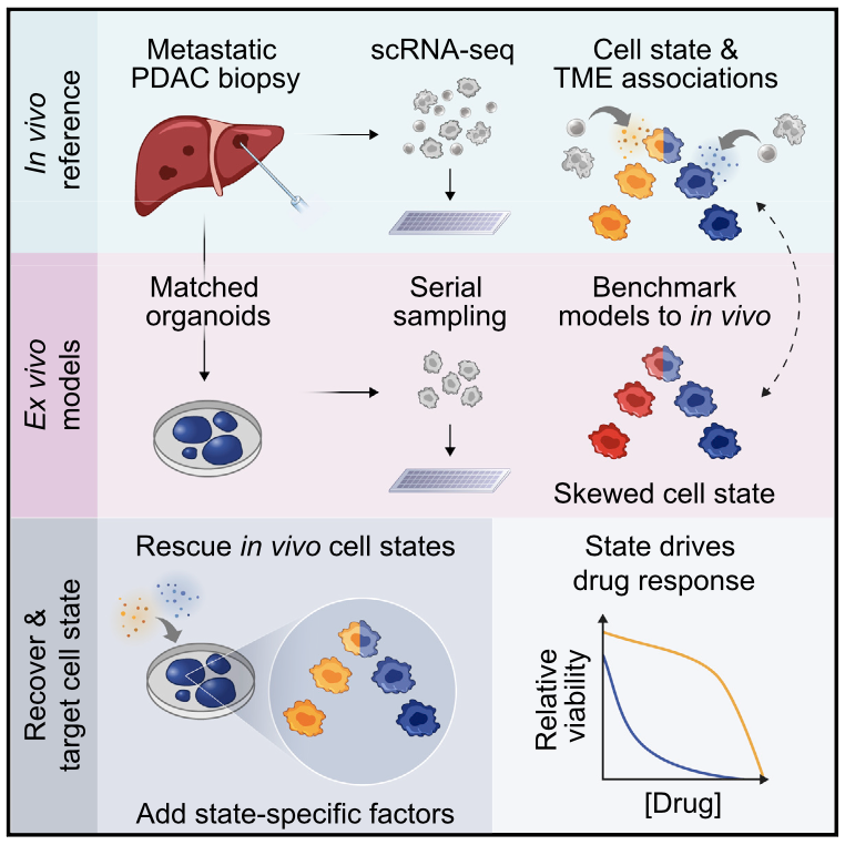
Prognostically relevant RNA expression states exist in pancreatic ductal adenocarcinoma (PDAC), but our understanding of their drivers, stability, and relationship to therapeutic response is limited. To examine these attributes systematically, we profiled metastatic biopsies and matched organoid models at single-cell resolution. In vivo, we identify a new intermediate PDAC transcriptional cell state and uncover distinct site- and state-specific tumor microenvironments (TMEs). Benchmarking models against this reference map, we reveal strong culture-specific biases in cancer cell transcriptional state representation driven by altered TME signals. We restore expression state heterogeneity by adding back in vivo-relevant factors and show plasticity in culture models. Further, we prove that non-genetic modulation of cell state can strongly influence drug responses, uncovering state-specific vulnerabilities. This work provides a broadly applicable framework for aligning cell states across in vivo and ex vivo settings, identifying drivers of transcriptional plasticity and manipulating cell state to target associated vulnerabilities.

Many patients infected with coronaviruses, such as SARS-CoV-2 and NL63 that use ACE2 receptors to infect cells, exhibit gastrointestinal symptoms and viral proteins are found in the human gastrointestinal tract, yet little is known about the inflammatory and pathological effects of coronavirus infection on the human intestine. Here, we used a human intestine-on-a-chip (Intestine Chip) microfluidic culture device lined by patient organoid-derived intestinal epithelium interfaced with human vascular endothelium to study host cellular and inflammatory responses to infection with NL63 coronavirus. These organoid-derived intestinal epithelial cells dramatically increased their ACE2 protein levels when cultured under flow in the presence of peristalsis-like mechanical deformations in the Intestine Chips compared to when cultured statically as organoids or in Transwell inserts. Infection of the intestinal epithelium with NL63 on-chip led to inflammation of the endothelium as demonstrated by loss of barrier function, increased cytokine production, and recruitment of circulating peripheral blood mononuclear cells (PBMCs). Treatment of NL63 infected chips with the approved protease inhibitor drug, nafamostat, inhibited viral entry and resulted in a reduction in both viral load and cytokine secretion, whereas remdesivir, one of the few drugs approved for COVID19 patients, was not found to be effective and it also was toxic to the endothelium. This model of intestinal infection was also used to test the effects of other drugs that have been proposed for potential repurposing against SARS-CoV-2. Taken together, these data suggest that the human Intestine Chip might be useful as a human preclinical model for studying coronavirus related pathology as well as for testing of potential anti-viral or anti-inflammatory therapeutics.

Leptomeningeal disease (LMD) is a devastating complication of solid tumor malignancies, with dire prognosis and no effective systemic treatment options. Over the past decade, the incidence of LMD has steadily increased due to therapeutics that have extended the survival of cancer patients, highlighting the need for new interventions. To examine the efficacy of immune checkpoint inhibitors (ICI) in patients with LMD, we completed two phase II clinical trials. Here, we investigate the cellular and molecular features underpinning observed patient trajectories in these trials by applying single-cell RNA and cell-free DNA profiling to longitudinal cerebrospinal fluid (CSF) draws from enrolled patients. We recover immune and malignant cell types in the CSF, characterize cell behavior changes following ICI, and identify genomic features associated with relevant clinical phenomena. Overall, our study describes the liquid LMD tumor microenvironment prior to and following ICI treatment and demonstrates clinical utility of cell-free and single-cell genomic measurements for LMD research.

Crohn’s disease is an inflammatory bowel disease (IBD) which most often presents with patchy lesions in the terminal ileum and colon and requires complex clinical care. Recent advances in the targeting of cytokines and leukocyte migration have greatly advanced treatment options, but most patients still relapse and inevitably progress. Although single-cell approaches are transforming our ability to understand the barrier tissue biology of inflammatory disease, comprehensive single-cell RNA-sequencing (scRNA-seq) atlases of IBD to date have largely sampled pre-treated patients with established disease. This has limited our understanding of which cell types, subsets, and states at diagnosis are predictive of disease severity and response to treatment. Here, through a combined clinical, flow cytometric, and scRNA-seq study, we profile diagnostic human biopsies from the terminal ileum of treatment-naive pediatric patients with Crohn’s disease (pediCD; n=14) and from non-inflamed pediatric controls with functional gastrointestinal disorders (FGID; n=13). To fully resolve and annotate epithelial, stromal, and immune cell states among the 201,883 single-cell transcriptomes, we develop and deploy a principled and unbiased tiered clustering approach, ARBOL, yielding 138 FGID and 305 pediCD end cell clusters. Notably, through both flow cytometry and scRNA-seq, we observe that at the level of broad cell types, treatment-naive pediCD is not readily distinguishable from FGID in cellular composition. However, by integrating high-resolution scRNA-seq analysis, we identify significant differences in cell states that arise during pediCD relative to FGID. Furthermore, by closely linking our scRNA-seq analysis with clinical meta-data, we resolve a vector of lymphoid, myeloid, and epithelial cell states in treatment-naive samples which can distinguish patients with less severe disease (those not on anti-TNF therapies (NOA)), from those with more severe disease at presentation who require anti-TNF therapies. Moreover, this vector was also able to distinguish those patients that achieve a full response (FR) to anti-TNF blockade from those more treatment-resistant patients who only achieve a partial response (PR). Our study jointly leverages a treatment-naive cohort, high-resolution principled scRNA-seq data analysis, and clinical outcomes to understand which baseline cell states may predict inflammatory disease trajectory.

Immune checkpoint blockers (ICBs) have failed in all phase III glioblastoma (GBM) trials. Here, we show that regulatory T (Treg) cells play a key role in GBM resistance to ICBs in experimental gliomas. Targeting glucocorticoid-induced TNFR-related receptor (GITR) in Treg cells using an agonistic antibody (αGITR) promotes CD4 Treg cell differentiation into CD4 effector T cells, alleviates Treg cell-mediated suppression of anti-tumor immune response, and induces potent anti-tumor effector cells in GBM. The reprogrammed GBM-infiltrating Treg cells express genes associated with a Th1 response signature, produce IFNγ, and acquire cytotoxic activity against GBM tumor cells while losing their suppressive function. αGITR and αPD1 antibodies increase survival benefit in three experimental GBM models, with a fraction of cohorts exhibiting complete tumor eradication and immune memory upon tumor re-challenge. Moreover, αGITR and αPD1 synergize with the standard of care treatment for newly-diagnosed GBM, enhancing the cure rates in these GBM models.

Intracerebral hemorrhage (ICH) is a devastating form of stroke with a high mortality rate and few treatment options. Discovery of therapeutic interventions has been slow given the challenges associated with studying acute injury in the human brain. Inflammation induced by exposure of brain tissue to blood appears to be a major part of brain tissue injury. Here, we longitudinally profiled blood and cerebral hematoma effluent from a patient enrolled in the Minimally Invasive Surgery with Thrombolysis in Intracerebral Hemorrhage Evacuation trial, offering a rare window into the local and systemic immune responses to acute brain injury. Using single-cell RNA-Seq (scRNA-Seq), this is the first report to our knowledge that characterized the local cellular response during ICH in the brain of a living patient at single-cell resolution. Our analysis revealed shifts in the activation states of myeloid and T cells in the brain over time, suggesting that leukocyte responses are dynamically reshaped by the hematoma microenvironment. Interestingly, the patient had an asymptomatic rebleed that our transcriptional data indicated occurred prior to detection by CT scan. This case highlights the rapid immune dynamics in the brain after ICH and suggests that sensitive methods such as scRNA-Seq would enable greater understanding of complex intracerebral events.

Opportunities to interrogate the immune responses in the injured tissue of living patients suffering from acute sterile injuries such as stroke and heart attack are limited. We leveraged a clinical trial of minimally invasive neurosurgery for patients with intracerebral hemorrhage (ICH), a severely disabling subtype of stroke, to investigate the dynamics of inflammation at the site of brain injury over time. Longitudinal transcriptional profiling of CD14+monocytes/macrophages and neutrophils from hematomas of patients with ICH revealed that the myeloid response to ICH within the hematoma is distinct from that in the blood and occurs in stages conserved across the patient cohort. Initially, hematoma myeloid cells expressed a robust anabolic proinflammatory profile characterized by activation of hypoxia-inducible factors (HIFs) and expression of genes encoding immune factors and glycolysis. Subsequently, inflammatory gene expression decreased over time, whereas anti-inflammatory circuits were maintained and phagocytic and antioxidative pathways up-regulated. During this transition to immune resolution, glycolysis gene expression and levels of the potent proresolution lipid mediator prostaglandin E2 remained elevated in the hematoma, and unexpectedly, these elevations correlated with positive patient outcomes. Ex vivo activation of human macrophages by ICH-associated stimuli highlighted an important role for HIFs in production of both inflammatory and anti-inflammatory factors, including PGE2, which, in turn, augmented VEGF production. Our findings define the time course of myeloid activation in the human brain after ICH, revealing a conserved progression of immune responses from proinflammatory to proresolution states in humans after brain injury and identifying transcriptional programs associated with neurological recovery.

Organ infiltration by donor T cells is critical to the development of acute graft-versus-host disease (aGVHD) in recipients after allogeneic hematopoietic stem cell transplant (allo-HCT). However, deconvoluting the transcriptional programs of newly recruited donor T cells from those of tissue-resident T cells in aGVHD target organs remains a challenge. Here, we combined the serial intravascular staining technique with single-cell RNA sequencing to dissect the tightly connected processes by which donor T cells initially infiltrate tissues and then establish a pathogenic tissue residency program in a rhesus macaque allo-HCT model that develops aGVHD. Our results enabled creation of a spatiotemporal map of the transcriptional programs controlling donor CD8+ T cell infiltration into the primary aGVHD target organ, the gastrointestinal (GI) tract. We identified the large and small intestines as the only two sites demonstrating allo-specific, rather than lymphodepletion-driven, T cell infiltration. GI-infiltrating donor CD8+ T cells demonstrated a highly activated, cytotoxic phenotype while simultaneously developing a canonical tissue-resident memory T cell (TRM) transcriptional signature driven by interleukin-15 (IL-15)/IL-21 signaling. We found expression of a cluster of genes directly associated with tissue invasiveness, including those encoding adhesion molecules (ITGB2), specific chemokines (CCL3 and CCL4L1) and chemokine receptors (CD74), as well as multiple cytoskeletal proteins. This tissue invasion transcriptional signature was validated by its ability to discriminate the CD8+ T cell transcriptome of patients with GI aGVHD from those of GVHD-free patients. These results provide insights into the mechanisms controlling tissue occupancy of target organs by pathogenic donor CD8+ TRMcells during aGVHD in primate transplant recipients.

Central to anti-tumor immunity are dendritic cells (DCs), which stimulate long-lived protective T cell responses. Recent studies have demonstrated that DCs can achieve a state of hyperactivation, which is associated with inflammasome activities within living cells. Herein, we report that hyperactive DCs have an enhanced ability to migrate to draining lymph nodes and stimulate potent cytotoxic T lymphocyte (CTL) responses. This enhanced migratory activity is dependent on the chemokine receptor CCR7 and is associated with a unique transcriptional program that is not observed in conventionally activated or pyroptotic DCs. We show that hyperactivating stimuli are uniquely capable of inducing durable CTL-mediated anti-tumor immunity against tumors that are sensitive or resistant to PD-1 inhibition. These protective responses are intrinsic to the cDC1 subset of DCs, depend on the inflammasome-dependent cytokine IL-1β, and enable tumor lysates to serve as immunogens. If these activities are verified in humans, hyperactive DCs may impact immunotherapy.

Understanding the earliest immune responses following HIV infection is critical to inform future vaccines and therapeutics. Here, we review recent prospective human studies in at-risk populations that have provided insight into immune responses during acute infection, including additional relevant data from non-human primate (NHP) studies. We discuss the timing, nature, and function of the diverse immune responses induced, the onset of immune dysfunction, and the effects of early anti-retroviral therapy administration. Treatment at onset of viremia mitigates peripheral T and B cell dysfunction, limits seroconversion, and enhances cellular antiviral immunity despite persistence of infection in lymphoid tissues. We highlight pertinent areas for future investigation, and how application of high-throughput technologies, alongside targeted NHP studies, may elucidate immune response features to target in novel preventions and cures.

Intracerebral hemorrhage (ICH) is a devastating form of stroke with a high mortality rate and few treatment options. Discovery of therapeutic interventions has been slow given the challenges associated with studying acute injury, particularly over time, in the human brain. Inflammation induced by exposure of brain tissue to blood appears to be a major part of brain tissue injury. Here we longitudinally profiled blood and cerebral hematoma effluent from a patient enrolled in the Minimally Invasive Surgery with Thrombolysis in Intracerebral Haemorrhage Evacuation (MISTIEIII) trial, offering a rare window into the local and systemic immune responses to acute brain injury. Using single-cell RNA-sequencing, we characterized the local cellular response during ICH in the brain of a living patient at single-cell resolution for the first time. Our analysis revealed rapid shifts in the activation states of myeloid and T cells in the brain over time, suggesting that leukocyte responses are dynamically reshaped by the hematoma microenvironment. Interestingly, the patient had an asymptomatic re-bleed (second local exposure to blood) that our transcriptional data indicated occurred more than 30 hours prior to detection by CT scan. This case highlights the rapid immune dynamics in the brain after ICH and suggests that sensitive methods like scRNA-seq can inform our understanding of complex intracerebral events.

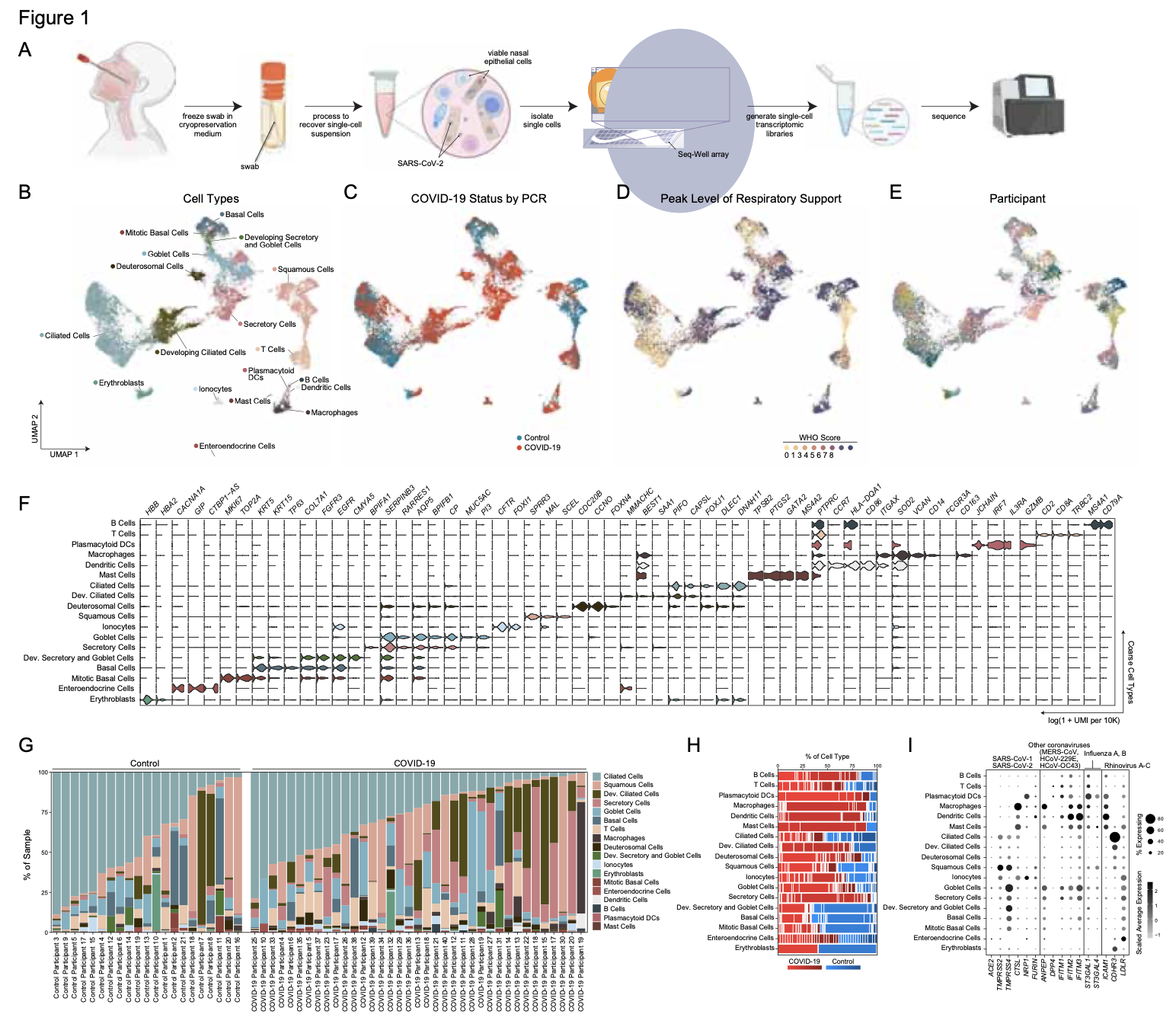
Our nasal epithelial COVID-19 dataset, along with COVID-19 datasets from other genomics groups, can now be found at covid19cellatlas.org. This work was sponsored by the Chan-Zuckerberg Initiative.

Bulk transcriptomic studies have defined classical and basal-like gene expression subtypes in pancreatic ductal adenocarcinoma (PDAC) that correlate with survival and response to chemotherapy; however, the underlying mechanisms that govern these subtypes and their heterogeneity remain elusive. Here, we performed single-cell RNA-sequencing of 23 metastatic PDAC needle biopsies and matched organoid models to understand how tumor cell-intrinsic features and extrinsic factors in the tumor microenvironment (TME) shape PDAC cancer cell phenotypes. We identify a novel cancer cell state that co-expresses basal-like and classical signatures, demonstrates upregulation of developmental and KRAS-driven gene expression programs, and represents a transitional intermediate between the basal-like and classical poles. Further, we observe structure to the metastatic TME supporting a model whereby reciprocal intercellular signaling shapes the local microenvironment and influences cancer cell transcriptional subtypes. In organoid culture, we find that transcriptional phenotypes are plastic and strongly skew toward the classical expression state, irrespective of genotype. Moreover, we show that patient-relevant transcriptional heterogeneity can be rescued by supplementing organoid media with factors found in the TME in a subtype-specific manner. Collectively, our study demonstrates that distinct microenvironmental signals are critical regulators of clinically relevant PDAC transcriptional states and their plasticity, identifies the necessity for considering the TME in cancer modeling efforts, and provides a generalizable approach for delineating the cell-intrinsic versus -extrinsic factors that govern tumor cell phenotypes.

Despite the epidemics of chronic obstructive pulmonary disease (COPD), the cellular and molecular mechanisms of this disease are far from being understood. Here, we characterize and classify the cellular composition within the alveolar space and peripheral blood of COPD patients and control donors using a clinically applicable single-cell RNA-seq technology corroborated by advanced computational approaches for: machine learning-based cell-type classification, identification of differentially expressed genes, prediction of metabolic changes, and modeling of cellular trajectories within a patient cohort. These high-resolution approaches revealed: massive transcriptional plasticity of macrophages in the alveolar space with increased levels of invading and proliferating cells, loss of MHC expression, reduced cellular motility, altered lipid metabolism, and a metabolic shift reminiscent of mitochondrial dysfunction in COPD patients. Collectively, single-cell omics of multi-tissue samples was used to build the first cellular and molecular framework for COPD pathophysiology as a prerequisite to develop molecular biomarkers and causal therapies against this deadly disease.

Barrier tissue epithelia play an essential role in maintaining organismal homeostasis, and changes in their cellular composition have been observed in multiple human diseases. Within the small intestinal epithelium, adult stem cells integrate diverse signals to regulate regeneration and differentiation, thereby establishing overall cellularity. Accordingly, directing stem cell differentiation could provide a tractable approach to alter the abundance or quality of specialized cells of the small intestinal epithelium, including the secretory Paneth, goblet, and enteroendocrine populations. Yet, to date, there has been a lack of suitable tools and rigorous approaches to identify biological targets and pharmacological agents that can modify epithelial composition to enable causal testing of disease-associated changes with novel therapeutic candidates. To empower the search for epithelia-modifying agents, we establish a first-of-its-kind high-throughput phenotypic organoid screen. We demonstrate the ability to screen thousands of samples and uncover biological targets and associated small molecule inhibitors which translate to in vivo. This approach is enabled by employing a functional, cell-type specific, scalable assay on an organoid model designed to represent the physiological cues of in vivo Paneth cell differentiation from adult intestinal stem cells. Further, we miniaturize and adapt the organoid culture system to enable automated plating and screening, thereby providing the ability to test thousands of samples. Strikingly, in our screen we identify inhibitors of the nuclear exporter Xpo1 modulate stem cell fate commitment by inducing a pan-epithelial stress response combined with an interruption of mitogen signaling in cycling intestinal progenitors, thereby significantly increasing the abundance of Paneth cells independent of known WNT and Notch differentiation cues. We extend our observation in vivo, demonstrating that oral administration of Xpo1 inhibitor KPT-330 at doses 1,000-fold lower than conventionally used in hematologic malignancies increases Paneth cell abundance. In total, we provide a framework to identify novel biological cues and therapeutic leads to rebalance intestinal stem cell differentiation and modulate epithelial tissue composition via high-throughput phenotypic screening in rationally-designed organoid model of differentiation.

Crucial transitions in cancer—including tumor initiation, local expansion, metastasis, and therapeutic resistance—involve complex interactions between cells within the dynamic tumor ecosystem. Transformative single-cell genomics technologies and spatial multiplex in situ methods now provide an opportunity to interrogate this complexity at unprecedented resolution. The Human Tumor Atlas Network (HTAN), part of the National Cancer Institute (NCI) Cancer Moonshot Initiative, will establish a clinical, experimental, computational, and organizational framework to generate informative and accessible three-dimensional atlases of cancer transitions for a diverse set of tumor types. This effort complements both ongoing efforts to map healthy organs and previous large-scale cancer genomics approaches focused on bulk sequencing at a single point in time. Generating single-cell, multiparametric, longitudinal atlases and integrating them with clinical outcomes should help identify novel predictive biomarkers and features as well as therapeutically relevant cell types, cell states, and cellular interactions across transitions. The resulting tumor atlases should have a profound impact on our understanding of cancer biology and have the potential to improve cancer detection, prevention, and therapeutic discovery for better precision-medicine treatments of cancer patients and those at risk for cancer.

There is pressing urgency to better understand the pathogenesis of the severe acute respiratory syndrome (SARS) coronavirus (CoV) clade SARS-CoV-2, which causes the disease known as COVID-19. SARS-CoV-2, like SARS-CoV, utilizes ACE2 to bind host cells. While initial SARS- CoV-2 cell entry and infection depend on ACE2 in concert with the protease TMPRSS2 for spike (S) protein activation, the specific cell subsets targeted by SARS-CoV-2 in host tissues, and the factors that regulate ACE2 expression, remain unknown. Here, we leverage human and non- human primate (NHP) single-cell RNA-sequencing (scRNA-seq) datasets to uncover the tissue- resident cell subsets that may serve as the cellular targets of SARS-CoV-2. We identify ACE2 and TMPRSS2 co-expressing cells within type II pneumocytes in NHP lung, absorptive enterocytes in human and NHP terminal ileum, and human nasal goblet secretory cells. Strikingly, we discover, and extensively corroborate using publicly available data sets, that ACE2 is an interferon-stimulated gene (ISG) in human epithelial cells. We further validate this finding in primary upper airway human respiratory epithelial cells. Thus, SARS-CoV-2 may exploit IFN- driven upregulation of ACE2, a key tissue-protective mediator during lung injury, to enhance infection.

Overview

In light of the global effort to better understand the new SARS-CoV-2 virus, we and other researchers from the HCA Lung Biological Network and beyond have begun an initiative to investigate datasets from relevant tissues profiled as part of other ongoing studies. These studies represent, for example, large efforts to characterize HIV, Mtb, and influenza infection and allergy in primary human and non-human primate samples. This page serves as a guide to viewing our data interactively on and downloading datasets from our single-cell portal, the Alexandria Project. Alternatively, bulk downloading of our data is available here. For more on research initiatives in COVID-19 being undertaken by the HCA, and the HCA Lung Biological Network in particular, please visit the HCA website here.

We investigated two genes whose protein products are central to the cellular entry of SARS-CoV-2: ACE2 and TMPRSS2. Consistent with previous studies, we found that the gene encoding ACE2, the SARS-CoV-2 entry receptor, is expressed on a subset of lung epithelial cells, type 2 pneumocytes, and a subset of ileal epithelial cells, absorptive enterocytes, across several datasets. The protease TMPRSS2 primes the spike protein of SARS-CoV-2 and is also important for viral entry. Because of this, we identified cells which co-express ACE2 and TMPRSS2 in our datasets, and investigated additional genes enriched within ACE2 and TMPRSS2 co-expressing cells. As we believe this data may prove useful to other researchers investigating similar questions, we have made our datasets public through the interactive Alexandria Project. Here, you can view our annotations of these datasets and investigate which other genes are highly expressed in these cell subsets of interest.

NB None of the datasets presented here were designed to answer specific questions about COVID-19. Additional studies will be required across larger, appropriately structured cohorts. Further, we provide a note of caution when interpreting scRNA-seq data for low abundance transcripts like ACE2 and TMPRSS2 as detection inefficiencies and/or sequencing depth may result in an underestimation of the actual frequencies of ACE2+ or ACE2+/TMPRSS2+ cells in tissues. Moreover, the protein levels of each may differ from their mRNA abundances. We present each data set separately, as each study differed by method of tissue processing and collection protocols, each of which can influence the frequency of recovered cell subsets.

Analysis

Our pre-print “SARS-CoV-2 receptor ACE2 is an interferon-stimulated gene in human airway epithelial cells and is enriched in specific cell subsets across tissues” can be found here, and the abstract is reproduced below.

There is pressing urgency to better understand the pathogenesis of the severe acute respiratory syndrome (SARS) coronavirus (CoV) clade SARS-CoV-2. SARS-CoV-2, like SARS-CoV, utilizes ACE2 to bind host cells. While initial SARS-CoV-2 cell entry and infection depend on ACE2 in concert with the protease TMPRSS2 for spike (S) protein activation, the specific cell subsets targeted by SARS-CoV-2 in host tissues, and the factors that regulate ACE2 expression, remain unknown. Here, we leverage human and non-human primate (NHP) single-cell RNA-sequencing (scRNA-seq) datasets to uncover the cell subsets that may serve as cellular targets of SARS-CoV-2. We identify ACE2/TMPRSS2 co-expressing cells within type II pneumocytes, absorptive enterocytes, and nasal goblet secretory cells. Strikingly, we discover that ACE2 is an interferon-stimulated gene (ISG) in human barrier tissue epithelial cells. Thus, SARS-CoV-2 may exploit IFN-driven upregulation of ACE2, a key tissue-protective mediator during lung injury, to enhance infection.

Datasets

Atlas of ACE2 expression in healthy non-human primate lung and ileum

In this study, we collected cells from various tissues in healthy and SHIV-infected non-human primates using Seq-Well v1. Here we highlight the lung and ileum and show that ACE2 and TMPRSS2 are co-expressed most frequently in type II pneumocytes in the lung and absorptive enterocytes in the ileum.

To visualize these cells in the Alexandria Project, visit this study: Atlas of healthy non-human primate lung and ileum ACE2+ cells

This project contains two UMAP visualizations (one for lung cells and one for ileum cells): toggle between them by clicking on the ‘Explore’ tab, select ‘View Options’ in the top right hand corner, and switch between lung and ileum under the ‘Load Cluster’ dropdown.

For each of the lung and ileum cell UMAPs, we provide subsets of these visualizations which contain only the cell types enriched for double positive cells. By selecting the ‘Load cluster’ options called ‘Lung Epithelial Cells’ or ‘Ileum Absorptive Enterocytes’, you will be able to view gene expression differences between double positive cells and other cells within those cell types. In the lung epithelial cells visualization, the following additional annotations are available under the ‘Select Annotation’ dropdown:

  • ACE2+ – cells expressing ACE2
  • TMPRSS2+ – cells expressing TMPRSS2
  • ACE2_TMPRSS2_double_positive – cells expressing both genes
  • celltype_double_positive – the cell type column and double positive column combined so genes that are differentially expressed between only one cell type may be viewed
  • celltype_ACE2+ – same as above for ACE2 expression

After selecting one of these annotations, you can then search for a gene of interest in the ‘Search Genes’ box in the left corner to view the expression of that gene as a violin plot split by the annotation you select. For example, to reproduce Figure 1D in the manuscript, you would select annotation “ACE2_TMPRSS2_double_positive” and search for gene ‘IFNGR2’ in the ‘Search Genes’ box.

The same options are available for the ileal absorptive enterocytes visualization except that the cell type is combined with the double positive/ACE2 column since this subset only includes one cell type.

Full expression matrices (and therefore gene expression values visible when using the ‘Search Genes’ box) are available only for epithelial cell types as this data is derived from a pre-publication study.

Atlas of ACE2 and TMPRSS2 in human ileum

In this study, samples from human ileum were collected, processed, and run on 10x 3′ v2 show ACE2 and TMPRSS2 co-expression in absorptive enterocytes.

To visualize these cells in the Alexandria Portal, visit this study: ACE2 and TMPRSS2 most enriched within GSTA1+MGST3+ absorptive enterocytes in context of non-inflamed terminal ileum

Two tSNE visualizations are available for this study, one of all cells in the ileum samples “non-inflammed-tsne” and one of just the epithelial cells in the samples “non-inflammed-epth-tsne”. To toggle between these, select the “Explore” tab under the study title, then select ‘View options’ from the upper right corner of the plot and choose the visualization of interest in the ‘Load cluster’ dropdown.

Full expression matrices (and therefore gene expression values visible when using the ‘search genes’ box) are available only for the epithelial cell types as this data is derived from a pre-publication study.

Atlas of ACE2 and TMPRSS2 expression in human HIV- and TB-infected lung

In this study, samples from lung surgeries were run with Seq-Well S^3 and contain a variety of immune and epithelial cell types. We found the majority of ACE2 and TPRSS2 double positive cells in type II pneumocyte cells.

To view these cells in the Alexandria Project, visit this study: Human lung HIV-TB co-infection ACE2+ cells

To visualize the double positive cells in these samples, click the ‘Explore’ tab, then select ‘View Options’ in the top right corner of the plot and choose ‘ACE2_TMPRSS2_double_positive’ under the ‘Select Annotation’ dropdown menu.

Full expression matrices (and therefore gene expression values visible when using the ‘search genes’ box) are available only for epithelial cell types as this data is derived from a pre-publication study.

A subset of ACE2+ secretory cells in human nasal mucosa

In this study, we find a subset of secretory cells which co-express ACE2 and TMPRSS2.

To visualize these cells on the Alexandria Project, visit this study: Allergic inflammatory memory in human respiratory epithelial progenitor cells

To view the tSNE of epithelial cells, select the ‘Explore’ tab, select ‘View Options’ in the right hand corner of the plot and choose ‘Epithelial cells’ in the ‘Load Cluster’ dropdown. To view the cell type annotations in the first panel of the above plot choose ‘subset’ in the ‘Select Annotation’ dropdown menu. To view the ACE2/TMPRSS2 double positive cells, select ‘ACE2_TMPRSS2’ from the ‘Select annotation dropdown menu, and to view the cluster subsets from the third panel of this plot, select ‘res_0_8’ from the ‘Select Annotation Dropdown Menu’.

ACE2 and TPRSS2 co-expressing cells found in non-human primate granulomas and adjacent uninvolved lung tissue

In this study, we collected lung tissue from non-human primates infected with mTB. These tissues come from both mTB granulomas and adjacent uninvolved lung in the same monkey.

These cells, profiled using Seq-Well S^3, can be investigated interactively in the Alexandria Project: Epithelial cells in NHP TB granuloma and uninvolved lung

To view the data colored by granuloma and uninvolved lung choose the “Granuloma” annotation accessible by clicking “View Options” in the top right-hand corner and selecting from the “Select Annotation” dropdown menu.

Full expression matrices (and therefore gene expression values visible when using the ‘Search Genes’ box) are available only for relevant cell types as this data is derived from a pre-publication study.

Comparison of ACE2 and TMPRSS2 expression in human duodenal and ileal tissue and organoid-derived epithelial cells

In this study, samples from adult human duodenum and ileum were collected and split for primary tissue single-cell RNA-seq and organoid culture under several conditions and profiled with Seq-Well S^3. Organoids were cultured and passaged every 6-8 days in Matrigel domes with established media conditions meant to recapitulate the broad diversity of in vivo epithelial cell types (Fujii, M., et al., Cell Stem Cell. 2018). Organoid culture media contained recombinant Noggin, Rspondin-3, FGF2, IGF1, afamin-Wnt3A, in addition to Gastrin and TGF-b inhibitor A83-01 with and without recombinant EGF (E/NR3+F2I1Gi+Af-W3+A83).  Cells co-expressing ACE2 and TMPRSS2 were identified principally within enterocyte clusters of both tissues and organoids.

Visualize these samples interactively and read more about this study on the Alexandria Project: Comparison of ACE2 and TMPRSS2 expression in human duodenal and ileal tissue and organoid-derived epithelial cells

This project contains two UMAP visualizations (one for organoid cells and one for primary tissue cells): toggle between them by clicking on the ‘Explore’ tab, select ‘View Options’ in the top right hand corner, and switch between tissue and organoid under the ‘Load Cluster’ dropdown.

To visualize cells which co-express ACE2 and TMPRSS2, select the ‘ACE2_TMPRSS2’ option under the ‘Select Annotation’ dropdown. You can then search for a gene of interest in the ‘Search Genes’ box in the left corner to view the expression of that gene as a violin plot split by the annotation you select.

Full expression matrices (and therefore gene expression values visible when using the ‘Search Genes’ box) are available only for enterocyte cell types as this data is derived from a pre-publication study. Expression of ACE2 and TMPRSS2 are available for all cells.

Interferon regulation of ACE2 in human and murine basal cells

Analysis of these datasets and others lead to the hypothesis that expression of the ACE2 receptor may be upregulated by interferon. To further interrogate this hypothesis, we cultured basal cells from two primary human donors, one human basal cell line, and one mouse trachea and stimulated them with  IL4, IL17a, IFNgamma, IFNαlpha, IFNbeta for 12 hours overnight. We performed bulk RNA sequencing and differential expression to show a dose dependent upregulation of canonical ISGs (interferon signaling genes). Specifically, we see that ACE2 is most significantly unregulated following IFN alpha stimulation in primary human basal cells, diminished in the BEAS-2B cell line and not seen in mouse cells. 

To visualize these samples, the gene expression data may be viewed interactively in the Alexandria Project: Interferon regulation of ACE2 in human and murine basal cells

Under the ‘Explore’ tab, use the ‘Search genes’ field in the top left corner to visualize log-normalized gene expression. To visualize expression in each sample, select ‘View Options’ in the top left corner of the plot and choose the sample of interest under ‘Load cluster’. The ‘Stim_Dose’ annotation refers to the dose of each stimulation condition applied to that sample. 

Data for all samples in this study can be dowloaded under the ‘Download’ tab.

Murine nasal mucosa after intranasal interferon exposure

We test the impact of IFNalpha stimulation in vivo by treating two mice intranasally with 200 ng of IFNalpha and two with saline. After 12 hours, the nasal mucosa of the respiratory and olfactory epithelia and underlying lamina propria were isolated and prepared for sequencing with Seq-Well S^3.

These cells may be viewed interactively in the Alexandria Project: Murine nasal mucosa after intranasal interferon exposure

Data for all samples in this study can be dowloaded under the ‘Download’ tab.

Alexandria Project Details

Alexandria Documentation

Single Cell Portal Documentation

We thank the Broad Institute Single Cell Portal team for creating the platform that allows Alexandria to exist and for their working tirelessly to help us share our datasets for others to access.

The induction of broadly neutralizing antibodies (bnAbs) is highly desired for an effective vaccine against HIV-1. Typically, bnAbs develop in patients with high viremia, but they can also evolve in some untreated HIV-1 controllers with low viral loads. Here, we identify a subgroup of neutralizer-controllers characterized by myeloid DCs (mDCs) with a distinct inflammatory signature and a superior ability to prime T follicular helper (Tfh)-like cells in an STAT4-dependent fashion. This distinct immune profile is associated with a higher frequency of Tfh-like cells in peripheral blood (pTfh) and an enrichment for Tfh-defining genes in circulating CD4+ T cells. Correspondingly, monocytes from this neutralizer controller subgroup upregulate genes encoding for chemotaxis and inflammation, and they secrete high levels of IL-12 in response to TLR stimulation. Our results suggest the existence of multi-compartment immune networks between mDCs, Tfh, and monocytes that may facilitate the development of bnAbs in a subgroup of HIV-1 controllers.

Mycobacterium tuberculosis (Mtb) is the leading cause of death from infection worldwide. The only available vaccine, BCG (Bacillus Calmette–Guérin), is given intradermally and has variable efficacy against pulmonary tuberculosis, the major cause of mortality and disease transmission. Here we show that intravenous administration of BCG profoundly alters the protective outcome of Mtb challenge in non-human primates (Macaca mulatta). Compared with intradermal or aerosol delivery, intravenous immunization induced substantially more antigen-responsive CD4 and CD8 T cell responses in blood, spleen, bronchoalveolar lavage and lung lymph nodes. Moreover, intravenous immunization induced a high frequency of antigen-responsive T cells across all lung parenchymal tissues. Six months after BCG vaccination, macaques were challenged with virulent Mtb. Notably, nine out of ten macaques that received intravenous BCG vaccination were highly protected, with six macaques showing no detectable levels of infection, as determined by positron emission tomography–computed tomography imaging, mycobacterial growth, pathology and granuloma formation. The finding that intravenous BCG prevents or substantially limits Mtb infection in highly susceptible rhesus macaques has important implications for vaccine delivery and clinical development, and provides a model for defining immune correlates and mechanisms of vaccine-elicited protection against tuberculosis.

Genome-wide association studies (GWAS) have identified genetic variants associated with age-related macular degeneration (AMD), one of the leading causes of blindness in the elderly. However, it has been challenging to identify the cell types associated with AMD given the genetic complexity of the disease. Here we perform massively parallel single-cell RNA sequencing (scRNA-seq) of human retinas using two independent platforms, and report the first single-cell transcriptomic atlas of the human retina. Using a multi-resolution network-based analysis, we identify all major retinal cell types, and their corresponding gene expression signatures. Heterogeneity is observed within macroglia, suggesting that human retinal glia are more diverse than previously thought. Finally, GWAS-based enrichment analysis identifies glia, vascular cells, and cone photoreceptors to be associated with the risk of AMD. These data provide a detailed analysis of the human retina, and show how scRNA-seq can provide insight into cell types involved in complex, inflammatory genetic diseases.

Antibody paratopes are formed by hypervariable complementarity-determining regions (CDRH3s) and variable gene-encoded CDRs. The latter show biased usage in human broadly neutralizing antibodies (bnAbs) against both HIV and influenza virus, suggesting the existence of gene-endowed targeting solutions that may be amenable to pathway amplification. To test this, we generated transgenic mice with human CDRH3 diversity but simultaneously constrained to individual user-defined human immunoglobulin variable heavy-chain (VH) genes, including IGHV1-69, which shows biased usage in human bnAbs targeting the hemagglutinin stalk of group 1 influenza A viruses. Sequential immunization with a stalk-only hemagglutinin nanoparticle elicited group 1 bnAbs, but only in IGHV1-69 mice. This VH-endowed response required minimal affinity maturation, was elicited alongside pre-existing influenza immunity, and when IGHV1-69 B cells were diluted to match the frequency measured in humans. These results indicate that the human repertoire could, in principle, support germline-encoded bnAb elicitation using a single recombinant hemagglutinin immunogen.

Pilocytic astrocytoma (PA), the most common childhood brain tumor, is a low-grade glioma with a single driver BRAF rearrangement. Here, we perform scRNAseq in six PAs using methods that enabled detection of the rearrangement. When compared to higher-grade gliomas, a strikingly higher proportion of the PA cancer cells exhibit a differentiated, astrocyte-like phenotype. A smaller proportion of cells exhibit a progenitor-like phenotype with evidence of proliferation. These express a mitogen-activated protein kinase (MAPK) programme that was absent from higher-grade gliomas. Immune cells, especially microglia, comprise 40% of all cells in the PAs and account for differences in bulk expression profiles between tumor locations and subtypes. These data indicate that MAPK signaling is restricted to relatively undifferentiated cancer cells in PA, with implications for investigational therapies directed at this pathway.

Genomic medicine has paved the way for identifying biomarkers and therapeutically actionable targets for complex diseases, but is complicated by the involvement of thousands of variably expressed genes across multiple cell types. Single-cell RNA-sequencing study (scRNA-seq) allows the characterization of such complex changes in whole organs. The study is based on applying network tools to organize and analyze scRNA-seq data from a mouse model of arthritis and human rheumatoid arthritis, in order to find diagnostic biomarkers and therapeutic targets. Diagnostic validation studies were performed using expression profiling data and potential protein biomarkers from prospective clinical studies of 13 diseases. A candidate drug was examined by a treatment study of a mouse model of arthritis, using phenotypic, immunohistochemical, and cellular analyses as read-outs. We performed the first systematic analysis of pathways, potential biomarkers, and drug targets in scRNA-seq data from a complex disease, starting with inflamed joints and lymph nodes from a mouse model of arthritis. We found the involvement of hundreds of pathways, biomarkers, and drug targets that differed greatly between cell types. Analyses of scRNA-seq and GWAS data from human rheumatoid arthritis (RA) supported a similar dispersion of pathogenic mechanisms in different cell types. Thus, systems-level approaches to prioritize biomarkers and drugs are needed. Here, we present a prioritization strategy that is based on constructing network models of disease-associated cell types and interactions using scRNA-seq data from our mouse model of arthritis, as well as human RA, which we term multicellular disease models (MCDMs). We find that the network centrality of MCDM cell types correlates with the enrichment of genes harboring genetic variants associated with RA and thus could potentially be used to prioritize cell types and genes for diagnostics and therapeutics. We validated this hypothesis in a large-scale study of patients with 13 different autoimmune, allergic, infectious, malignant, endocrine, metabolic, and cardiovascular diseases, as well as a therapeutic study of the mouse arthritis model. Overall, our results support that our strategy has the potential to help prioritize diagnostic and therapeutic targets in human disease.

Sustained viremia after acute HIV infection is associated with profound CD4+ T cell loss and exhaustion of HIV-specific CD8+ T cell responses. To determine the impact of combination antiretroviral therapy (cART) on these processes, we examined the evolution of immune responses in acutely infected individuals initiating treatment before peak viremia. Immediate treatment of Fiebig stages I and II infection led to a rapid decline in viral load and diminished magnitude of HIV-specific (tetramer+) CD8+ T cell responses compared to untreated donors. There was a strong positive correlation between cumulative viral antigen exposure before full cART-induced suppression and immune responses measured by MHC class I tetramers, IFN-γ ELISPOT, and CD8+ T cell activation. HIV-specific CD8+ T responses of early treated individuals were characterized by increased CD127 and BCL-2 expression, greater in vitro IFN-γ secretion, and enhanced differentiation into effector memory (Tem) cells. Transcriptional analysis of tetramer+ CD8+ T cells from treated persons revealed reduced expression of genes associated with activation and apoptosis, with concurrent up-regulation of prosurvival genes including BCL-2AXL, and SRC. Early treatment also resulted in robust HIV-specific CD4+ T cell responses compared to untreated HIV-infected individuals. Our data show that limiting acute viremia results in enhanced functionality of HIV-specific CD4+ and CD8+ T cells, preserving key antiviral properties of these cells.

The liver can substantially regenerate after injury, with both main epithelial cell types, hepatocytes and biliary epithelial cells (BECs), playing important roles in parenchymal regeneration. Beyond metabolic functions, BECs exhibit substantial plasticity and in some contexts can drive hepatic repopulation. Here, we performed single-cell RNA sequencing to examine BEC and hepatocyte heterogeneity during homeostasis and after injury. Instead of evidence for a transcriptionally defined progenitor-like BEC cell, we found significant homeostatic BEC heterogeneity that reflects fluctuating activation of a YAPdependent program. This transcriptional signature defines a dynamic cellular state during homeostasis and is highly responsive to injury. YAP signaling is induced by physiological bile acids (BAs), required for BEC survival in response to BA exposure, and is necessary for hepatocyte reprogramming into biliary progenitors upon injury. Together, these findings uncover molecular heterogeneity within the ductal epithelium and reveal YAP as a protective rheostat and regenerative regulator in the mammalian liver.

In the small intestine, a niche of accessory cell types supports the generation of mature epithelial cell types from intestinal stem cells (ISCs). It is unclear, however, if and how immune cells in the niche affect ISC fate or the balance between self-renewal and differentiation. Here, we use single-cell RNA sequencing (scRNA-seq) to identify MHC class II (MHCII) machinery enrichment in two subsets of Lgr5 + ISCs. We show that MHCII +Lgr5 + ISCs are non-conventional antigen-presenting cells in co-cultures with CD4 + T helper (Th) cells. Stimulation of intestinal organoids with key Th cytokines affects Lgr5 + ISC renewal and differentiation in opposing ways: pro-inflammatory signals promote differentiation, while regulatory cells and cytokines reduce it. In vivo genetic perturbation of Th cells or MHCII expression on Lgr5 + ISCs impacts epithelial cell differentiation and IEC fate during infection. These interactions between Th cells and Lgr5 + ISCs, thus, orchestrate tissue-wide responses to external signals.

Genome-wide association studies (GWAS) have revealed risk alleles for ulcerative colitis (UC), but their cell type and pathway specificities are often unknown. Here, we generate an atlas of 115,517 cells from the colon mucosa of seven UC patients and ten healthy individuals, revealing 51 epithelial, stromal, and immune cell subsets, including a subset of BEST4+ enterocytes, which may sense and respond to pH, and IL13RA2+IL-11+ inflammatory fibroblasts, which we associate with resistance to anti-TNF therapy. Inflammatory fibroblasts, inflammatory monocytes, microfold-like cells, and CD8+IL-17+ T cells expand during disease, and form intercellular interaction hubs that mediate cross-talk between diverse cellular lineages. We identify hundreds of putative autocrine and paracrine cell-cell interactions that may explain the migration, expansion, or inhibition of cell types with disease. Surprisingly, UC risk genes are often cell type specific and co-regulated in relatively few gene modules, suggesting convergence onto limited sets of cell types and pathways. Using this observation, we nominate and infer putative functions for UC risk genes across all GWAS loci. Our atlas thus provides a framework for interrogating complex human diseases and mapping risk variants onto their cell types and pathways of activity.

Barrier tissue dysfunction is a fundamental feature of chronic human inflammatory diseases1. Specialized subsets of epithelial cells—including secretory and ciliated cells—differentiate from basal stem cells to collectively protect the upper airway2,3,4. Allergic inflammation can develop from persistent activation5 of type 2 immunity6 in the upper airway, resulting in chronic rhinosinusitis, which ranges in severity from rhinitis to severe nasal polyps7. Basal cell hyperplasia is a hallmark of severe disease7,8,9, but it is not known how these progenitor cells2,10,11contribute to clinical presentation and barrier tissue dysfunction in humans. Here we profile primary human surgical chronic rhinosinusitis samples (18,036 cells, n = 12) that span the disease spectrum using Seq-Well for massively parallel single-cell RNA sequencing12, report transcriptomes for human respiratory epithelial, immune and stromal cell types and subsets from a type 2 inflammatory disease, and map key mediators. By comparison with nasal scrapings (18,704 cells, n = 9), we define signatures of core, healthy, inflamed and polyp secretory cells. We reveal marked differences between the epithelial compartments of the non-polyp and polyp cellular ecosystems, identifying and validating a global reduction in cellular diversity of polyps characterized by basal cell hyperplasia, concomitant decreases in glandular cells, and phenotypic shifts in secretory cell antimicrobial expression. We detect an aberrant basal progenitor differentiation trajectory in polyps, and propose cell-intrinsic13, epigenetic14,15 and extrinsic factors11,16,17 that lock polyp basal cells into this uncommitted state. Finally, we functionally demonstrate that ex vivo cultured basal cells retain intrinsic memory of IL-4/IL-13 exposure, and test the potential for clinical blockade of the IL-4 receptor α-subunit to modify basal and secretory cell states in vivo. Overall, we find that reduced epithelial diversity stemming from functional shifts in basal cells is a key characteristic of type 2 immune-mediated barrier tissue dysfunction. Our results demonstrate that epithelial stem cells may contribute to the persistence of human disease by serving as repositories for allergic memories.

Background

Human immunity relies on the coordinated responses of many cellular subsets and functional states. Inter-individual variations in cellular composition and communication could thus potentially alter host protection. Here, we explore this hypothesis by applying single-cell RNA-sequencing to examine viral responses among the dendritic cells (DCs) of three elite controllers (ECs) of HIV-1 infection.

Results

To overcome the potentially confounding effects of donor-to-donor variability, we present a generally applicable computational framework for identifying reproducible patterns in gene expression across donors who share a unifying classification. Applying it, we discover a highly functional antiviral DC state in ECs whose fractional abundance after in vitro exposure to HIV-1 correlates with higher CD4+ T cell counts and lower HIV-1 viral loads, and that effectively primes polyfunctional T cell responses in vitro. By integrating information from existing genomic databases into our reproducibility-based analysis, we identify and validate select immunomodulators that increase the fractional abundance of this state in primary peripheral blood mononuclear cells from healthy individuals in vitro.

Conclusions

Overall, our results demonstrate how single-cell approaches can reveal previously unappreciated, yet important, immune behaviors and empower rational frameworks for modulating systems-level immune responses that may prove therapeutically and prophylactically useful.


Complete information about the scRAD R package is available on the Shalek Lab Resources page.

The recent advent of methods for high-throughput single-cell molecular profiling has catalyzed a growing sense in the scientific community that the time is ripe to complete the 150-year-old effort to identify all cell types in the human body. The Human Cell Atlas Project is an international collaborative effort that aims to define all human cell types in terms of distinctive molecular profiles (such as gene expression profiles) and to connect this information with classical cellular descriptions (such as location and morphology). An open comprehensive reference map of the molecular state of cells in healthy human tissues would propel the systematic study of physiological states, developmental trajectories, regulatory circuitry and interactions of cells, and also provide a framework for understanding cellular dysregulation in human disease. Here we describe the idea, its potential utility, early proofs-of-concept, and some design considerations for the Human Cell Atlas, including a commitment to open data, code, and community.

Tissue barrier dysfunction is a poorly defined feature hypothesized to drive chronic human inflammatory disease. The epithelium of the upper respiratory tract represents one such barrier, responsible for separating inhaled agents, such as pathogens and allergens, from the underlying submucosa. Specialized epithelial subsets-including secretory, glandular, and ciliated cells-differentiate from basal progenitors to collectively realize this role. Allergic inflammation in the upper airway barrier can develop from persistent activation of Type 2 immunity (T2I), resulting in the disease spectrum known as chronic rhinosinusitis (CRS), ranging from rhinitis to severe nasal polyps. Whether recently identified epithelial progenitor subsets, and their differentiation trajectory, contribute to the clinical presentation and barrier dysfunction in T2I-mediated disease in humans remains unexplored. Profiling twelve primary human CRS samples spanning the range of clinical severity with the Seq-Well platform for massively-parallel single-cell RNA-sequencing (scRNA-seq), we report the first single-cell transcriptomes for human respiratory epithelial cell subsets, immune cells, and parenchymal cells (18,036 total cells) from a T2I inflammatory disease, and map key mediators. We find striking differences between non-polyp and polyp tissues within the epithelial compartments of human T2I cellular ecosystems. More specifically, across 10,383 epithelial cells, we identify a global reduction in epithelial diversity in polyps characterized by basal cell hyperplasia, a concomitant decrease in glandular and ciliated cells, and phenotypic shifts in secretory cell function. We validate these findings through flow cytometry, histology, and bulk tissue RNA-seq of an independent cohort. Furthermore, we detect an aberrant basal progenitor differentiation trajectory in polyps, and uncover cell-intrinsic and extrinsic factors that may lock polyp basal cells into an uncommitted state. Overall, our data define severe T2I barrier dysfunction as a reduction in epithelial diversity, characterized by profound functional shifts stemming from basal cell defects, and nominate a cellular mechanism for the persistence and chronicity of severe human respiratory disease.

Click here to read the pre-publication manuscript.

Single-cell RNA-seq could play a key role in personalized medicine by facilitating characterization of cells, pathways, and genes associated with human diseases such as cancer.

We broadly study how intra- and extracellular circuits collectively drive healthy and diseased tissue states. By leveraging the massive genomic datasets we and others have generated from complex tissues (like melanoma tumors, inflamed gut, and nasal polyps), we have begun to identify common and unique cell types/states and circuits associated with pathology that may be important for regulating biological function and stability. Our current findings suggest multiple overlaps among distinct diseases, pointing to the possibility of a finite set of evolved response strategies and thus common interventions based on adjusting specific cell states, cell frequencies, and/or cell-cell communication pathways.

Vaccines remain the most effective tool to prevent infectious diseases. Here, we introduce an in vitro booster vaccination approach that relies on antigen-dependent activation of human memory B cells in culture. This stimulation induces antigen-specific B cell proliferation, differentiation of B cells into plasma cells, and robust antibody secretion after a few days of culture. We validated this strategy using cells from healthy donors to retrieve human antibodies against tetanus toxoid and influenza hemagglutinin (HA) from H1N1 and newly emergent subtypes such as H5N1 and H7N9. Anti-HA antibodies were cross-reactive against multiple subtypes, and some showed neutralizing activity. Although these antibodies may have arisen as a result of previous influenza infection, we also obtained gp120-reactive antibodies from non–HIV-infected donors, indicating that we can generate antibodies without prior antigenic exposure. Overall, our novel approach can be used to rapidly produce therapeutic antibodies and has the potential to assess the immunogenicity of candidate antigens, which could be exploited in future vaccine development.

We lack effective treatments and preventions for many of the most challenging infectious diseases, many of which disproportionately impact those in low- and middle-income countries or traditionally marginalized communities.

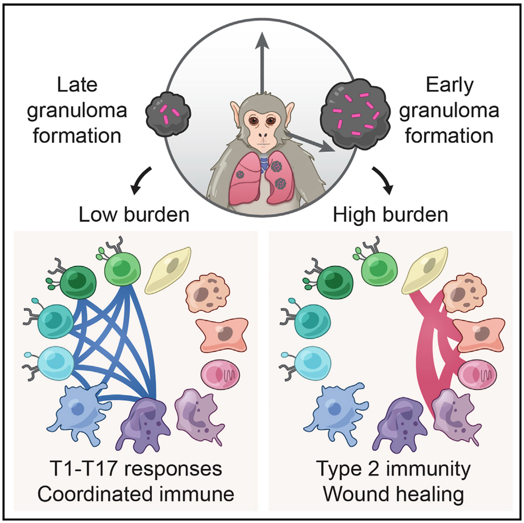
To help address this, we have established and enabled multi-group, multi-country partnerships to deploy and adapt cutting-edge genomic tools. By examining how cells dynamically alter their states, individually and collectively, during disease and/or its resolution in acute and chronic infections—e.g., tuberculosis, HIV/SHIV, hepatitis, malaria, leprosy, flu, SARS-CoV-2, and ebola—we have uncovered cellular and molecular features of pathogen control or pathology to potentiate or counteract, respectively. Illustratively, in tuberculosis, we identified a functional role for cytotoxic CD8 and hybrid type1-type17 T cells in control of infection in the lung and links between mast, plasma, and endothelial cell abundance (type-2 immune responses) and bacterial burden. We have also built methods for examining pathogens within individual host cells to define their dynamic interdependence and identify potentially restrictive host factors.

We are currently working to identify the drivers of common host responses to distinct perturbations and their targetability, as well as the impact of different interventions (e.g., vaccines).

Immune responses play a critical role in preventing tumorigenesis. Sometimes, however, they are ineffectual and can even drive/support malignancy.

We have examined how cancer cells alter and are influenced by their tumor microenvironments (TMEs), and the impact this has on therapeutic responses. Illustratively, in Pancreatic Ductal Adenocarcinoma (PDAC), by profiling liver metastases and matched organoid models, we showed: 1. associations between TME and malignant cell state composition; 2. that autocrine and paracrine signaling can drive malignant cell state transitions, even in an isogenic background, altering the efficacy of frontline chemotherapies; and, 3. that microenvironmental manipulations can be used to control malignant state, and thereby drug responses, rationally, and to improve model fidelity for screening potential therapies. This and related work highlight the potential utility of modulating indirect target cells (T cells in the PDAC TME or basal cells in allergic inflammation) to enhance cures and preventions. 

We are now systematically expanding this work to define how additional environmental and cell-intrinsic factors influence malignant cell state plasticity in PDAC and other cancers toward enhancing treatments.


We are exposed to a constant flux of external biochemical and physical stimuli as we age. Despite variability in our overall experiences and exact constitutions, our individual tissues typically manage to maintain functionality, though each can differ in its resilience to distinct stressors.

We have characterized how differences in cellular composition and communication impact tissue fitness and have identified responses and subsequent adaptations that drive chronic dysfunction. For example, although aberrant immune activity can precipitate allergic inflammatory diseases, therapies targeting immune cells and signaling are only successful in some, suggesting chronicity may involve alternative mechanisms. Previously, we helped demonstrate that dysregulated type-2 immune signaling, driven by environmental allergens, can impact tissue health in the upper airway through generating dysfunctional basal epithelial stem cells. These stem cells can then contribute to persistence by serving as repositories for allergic inflammatory memories, altering the integrity and functional output of the nasal epithelium. Our work, with that of others, suggests generalizable principles for cellular memory, and informs where and how tissues should be targeted to support health or restore function. We have since further investigated how tissue-resident cellular subsets participate in, and are shaped by, environmental exposures at barrier tissues and the functional consequences of these experiences.

We are now working to develop a more holistic appreciation for how different intra- and extracellular factors (e.g., genetics and integrated exposure history, respectively) influence barrier tissue function.

To explore the distinct genotypic and phenotypic states of melanoma tumors, we applied single-cell RNA sequencing (RNA-seq) to 4645 single cells isolated from 19 patients, profiling malignant, immune, stromal, and endothelial cells. Malignant cells within the same tumor displayed transcriptional heterogeneity associated with the cell cycle, spatial context, and a drug-resistance program. In particular, all tumors harbored malignant cells from two distinct transcriptional cell states, such that tumors characterized by high levels of the MITF transcription factor also contained cells with low MITF and elevated levels of the AXL kinase. Single-cell analyses suggested distinct tumor microenvironmental patterns, including cell-to-cell interactions. Analysis of tumor-infiltrating T cells revealed exhaustion programs, their connection to T cell activation and clonal expansion, and their variability across patients. Overall, we begin to unravel the cellular ecosystem of tumors and how single-cell genomics offers insights with implications for both targeted and immune therapies.