Alex Profiled in MIT News
Jul 22, 2022
Read Alex’s new profile in MIT News here.

The Shalek Lab develops and applies broadly applicable experimental and computational platforms to understand and engineer immune responses in tissues. We employ a comprehensive, five-step approach, building innovative methodologies and leveraging them with partners around the world to facilitate deeper, more mechanistic inquiry into how cells drive tissue-level behaviors across the spectrum of human health and disease.

 Biology
 Cell Atlas
 Genomics
 Immunology
 Infectious Disease
 Microbiology
 Alex K. Shalek
 Conner Kummerlowe
 Constantine Tzouanas
 José Ordovas-Montañes
 Marc Wadsworth II
 Samira Ibrahim
 Sarah Nyquist
 Travis Hughes
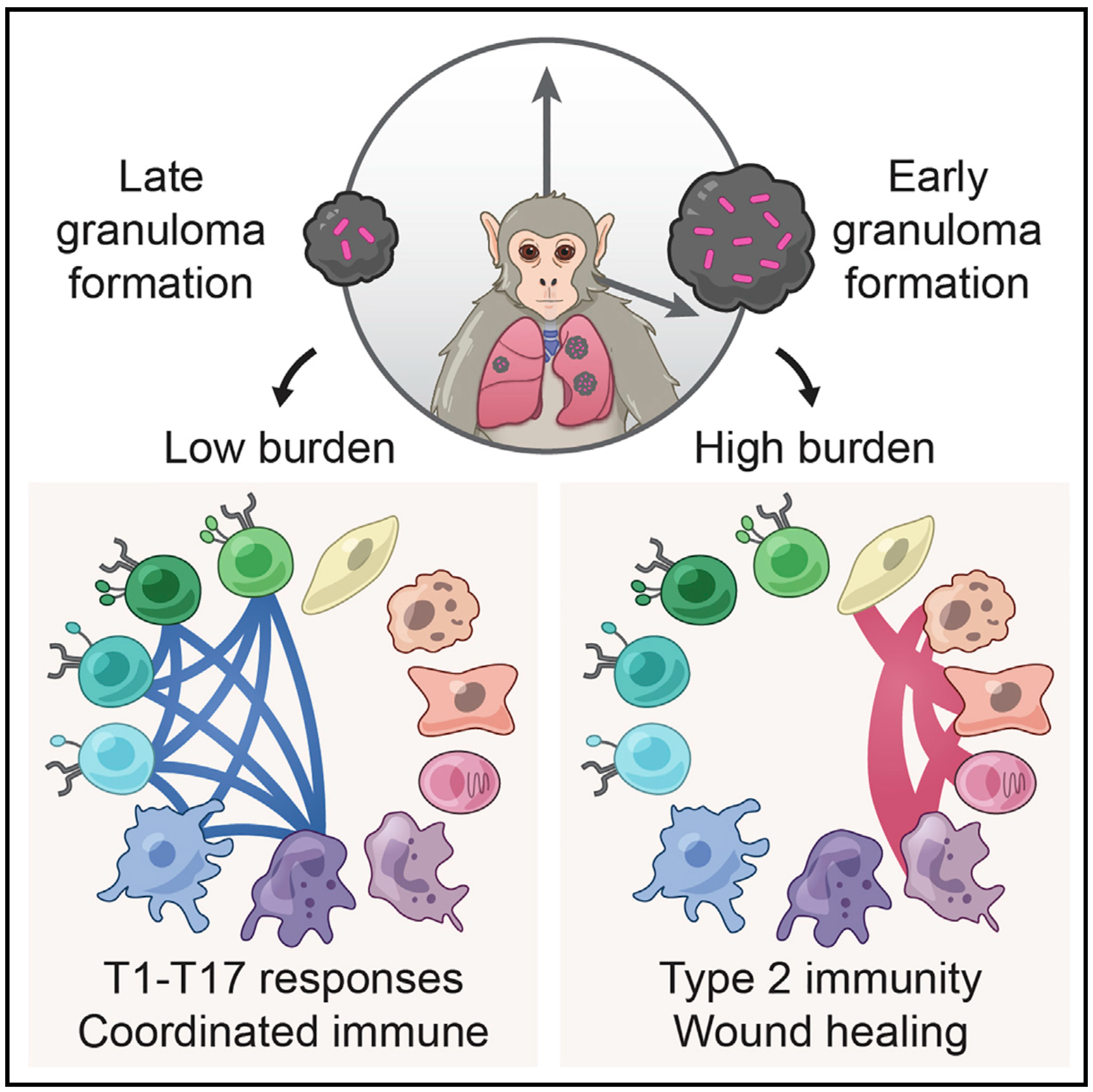
We integrate PET and CT imaging, scRNA-seq, and measures of bacterial clearance on TB granulomas from cynomolgus macaques to identify cellular features and programs associated with bacterial control and persistence of granulomas.
 Cancer
 Computational Methods
Genomics
Immunology
Computational Methods
Genomics
Immunology
 Medicine
 R&D
R&D
 Technology
Alex K. Shalek
Technology
Alex K. Shalek
 Andrew Navia
 Jennyfer Galvez-Reyes
 Nolawit Mulugeta
 Peter Winter
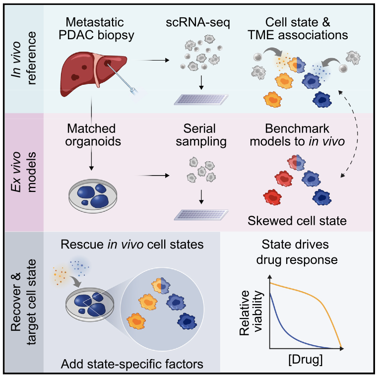
We profile metastatic biopsies and matched organoids to show that cancer cell state influences, and can be manipulated to shape, therapeutic response in pancreatic ductal adenocarcinoma.
 Biology
Cell Atlas
Computational Methods
Genomics
Immunology
Infectious Disease
Alex K. Shalek
Andrew Navia
Biology
Cell Atlas
Computational Methods
Genomics
Immunology
Infectious Disease
Alex K. Shalek
Andrew Navia
 Carly Ziegler
 Josh Bromley
José Ordovas-Montañes
Josh Bromley
José Ordovas-Montañes
 Micayla George
 Riley Drake
 Vincent Miao
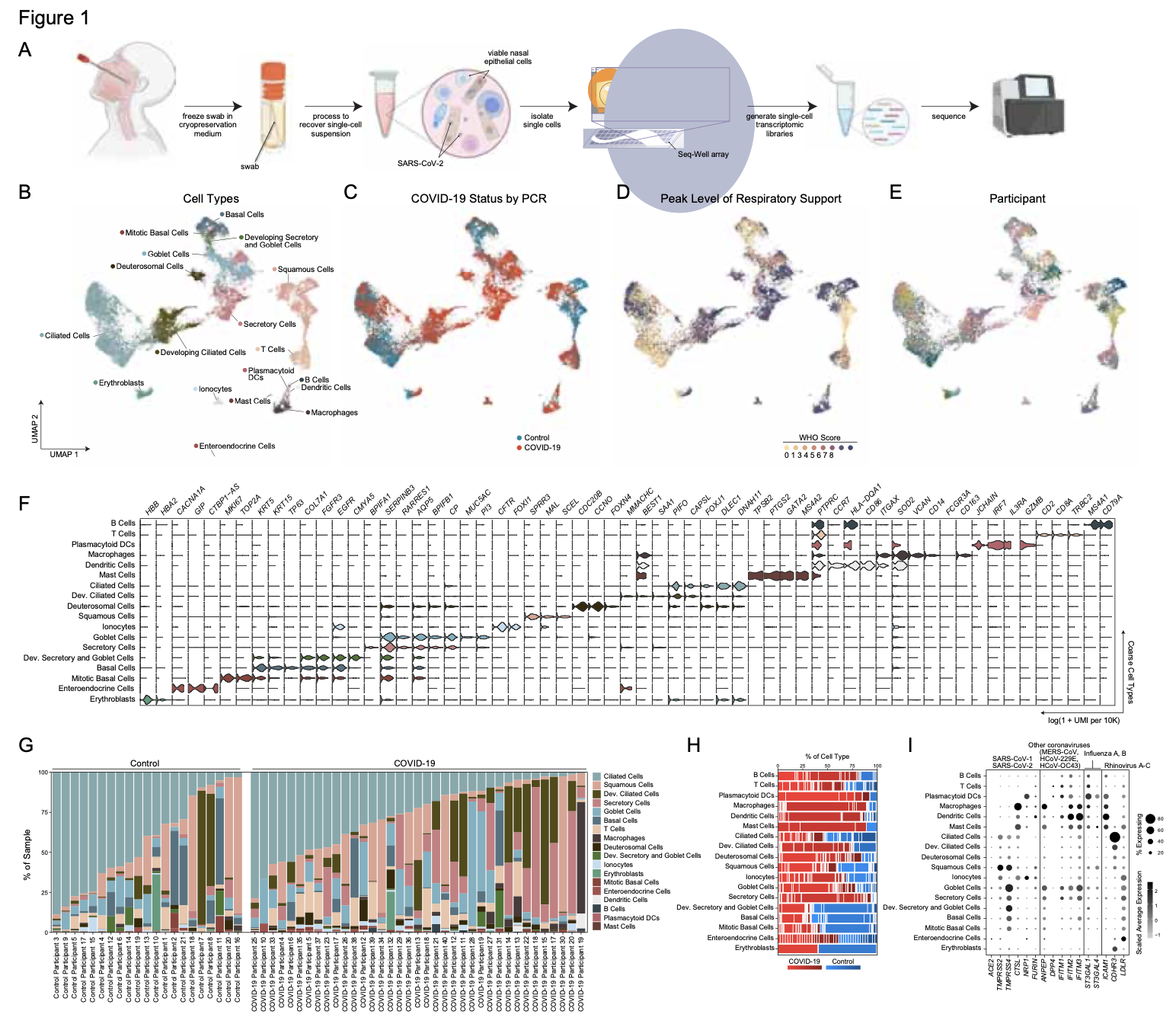
We analyze the nasopharyngeal cellular landscape of individuals with COVID-19, identifying putative initial target cells of SARS-CoV-2. Further, we find a blunted epithelial anti-viral response is correlated with the development of severe disease.
Jul 22, 2022
Read Alex’s new profile in MIT News here.
May 10, 2022
Check out the preview of our recent paper on TB granulomas here.
Mar 18, 2022
Listen to Alex Shalek on the Bioinformatics CRO Podcast Hear Alex talk about the Shalek Lab’s multi-disciplinary approach to answering some of science’s toughest and complex questions.