Computational cohort materials from March 2023 Ghana training
Hepatitis B virus (HBV) infection is restricted to the liver where it drives exhaustion of virus-specific T and B cells and pathogenesis through dysregulation of intrahepatic immunity. Our understanding of liver-specific events related to viral control and liver damage have relied almost solely on animal models and we lack useable peripheral biomarkers to quantify intrahepatic immune activation beyond cytokine measurement. Our objective was to overcome practical obstacles of liver sampling using fine-needle aspiration (FNA) and develop an optimized workflow to comprehensively compare the blood and liver compartments within chronic hepatitis B (CHB) patients using single-cell RNA sequencing (scRNAseq). We developed a workflow that enabled multi-site international studies and centralized scRNAseq. Blood and liver FNAs were collected, and cellular and molecular capture were compared between the Seq-Well S3 picowell-based and the 10x Chromium reverse-emulsion droplet-based scRNAseq technologies. Both technologies captured the cellular diversity of the liver but Seq-Well S3 effectively captured neutrophils, which were absent in the 10x dataset. CD8 T cells and neutrophils displayed distinct transcriptional profiles between blood and liver. In addition, liver FNAs captured a heterogeneous liver macrophage population. Comparison between untreated CHB patients and patients treated with nucleoside analogues showed that myeloid cells were highly sensitive to environmental changes while lymphocytes displayed minimal differences. The ability to electively sample and intensively profile the immune landscape of the liver, and generate high-resolution data, will enable multi-site clinical studies to identify biomarkers for intrahepatic immune activity in HBV and beyond.
Imidazoquinolines (IMDs), such as resiquimod (R848), are of great interest as potential cancer immunotherapies because of their ability to activate Toll-like receptor 7 (TLR7) and/or TLR8 on innate immune cells. Nevertheless, intravenous administration of IMDs causes severe immune-related toxicities, and attempts to improve their tissue-selective exposure while minimizing acute systemic inflammation have proven difficult. Here, using a library of R848 “bottlebrush prodrugs” (BPDs) that differ only by their R848 release kinetics, we explore how the timing of R848 exposure affects immune stimulation in vitro and in vivo. These studies led to the discovery of R848-BPDs that exhibit optimal activation kinetics to achieve potent stimulation of myeloid cells in tumors and substantial reductions in tumor growth following systemic administration in mouse syngeneic tumor models without any observable systemic toxicity. These results suggest that release kinetics can be tuned at the molecular level to provide safe yet effective systemically administered immunostimulant prodrugs for next-generation cancer immunotherapies.
Chronic liver disease and hepatocellular carcinoma (HCC) are life-threatening diseases with limited treatment options. The lack of clinically relevant/tractable experimental models hampers therapeutic discovery. Here, we develop a simple and robust human liver cell-based system modeling a clinical prognostic liver signature (PLS) predicting long-term liver disease progression toward HCC. Using the PLS as a readout, followed by validation in nonalcoholic steatohepatitis/fibrosis/HCC animal models and patient-derived liver spheroids, we identify nizatidine, a histamine receptor H2 (HRH2) blocker, for treatment of advanced liver disease and HCC chemoprevention. Moreover, perturbation studies combined with single cell RNA-Seq analyses of patient liver tissues uncover hepatocytes and HRH2+, CLEC5Ahigh, MARCOlowliver macrophages as potential nizatidine targets. The PLS model combined with single cell RNA-Seq of patient tissues enables discovery of urgently needed targets and therapeutics for treatment of advanced liver disease and cancer prevention.
Vitrification is a method for long-term biological sample cryopreservation that transforms cells into a glass-like state by cooling without causing intra- and extra-cellular ice formation, which is a major driver of cell cryoinjury. Compared to slow freezing, another conventional cryopreservation method, vitrification is simple, cost- effective and does not require a complex programmable freezer. Vitrification has been increasingly used to cryopreserve gametes and embryos for fertility preservation in assisted reproductive technology (ART). Moreover, vitrification of individual follicles followed by in vitro maturation (IVM) has emerged as a new fertility preservation method, particularly for childhood cancer patients who have no mature oocytes available for harvesting and for patients who cannot undergo ovarian tissue transplantation after cryopreservation because of the risk of reintroducing malignant cells. However, vitrification of individual follicles has been challenging because intact follicles have a more complex structure and larger size than individual oocytes or early embryos. Traditional oocyte/embryo vitrification methods are not optimized for individual follicles, and have been shown to compromise the qualities of follicles or oocytes, partly by damaging the gap junction between follicular cells or the transzonal projections (TZP) between the oocyte and cumulus cells. In our previous studies, we developed a closed vitrification method for cryopreserving ovarian tissues that was modified for individual follicles. Furthermore, using an alginate hydrogel encapsulated in vitro follicle growth (eIVFG) system, we have recently demonstrated that compared to freshly-harvested follicles, vitrified follicles have normal follicle and oocyte reproductive outcomes as well as comparable expression levels of several genes that are essential for gonadotropin-dependent folliculogenesis and oogenesis. However, it is unknown whether vitrification preserves the molecular signatures of folliculogenesis at the whole transcriptomic level, which is the primary research focus in this study.
High-throughput phenotypic screens leveraging biochemical perturbations, high-content readouts, and complex multicellular models could advance therapeutic discovery yet remain constrained by limitations of scale. To address this, we establish a method for compressing screens by pooling perturbations followed by computational deconvolution. Conducting controlled benchmarks with a highly bioactive small molecule library and a high-content imaging readout, we demonstrate increased efficiency for compressed experimental designs compared to conventional approaches. To prove generalizability, we apply compressed screening to examine transcriptional responses of patient-derived pancreatic cancer organoids to a library of tumor-microenvironment (TME)-nominated recombinant protein ligands. Using single-cell RNA-seq as a readout, we uncover reproducible phenotypic shifts induced by ligands that correlate with clinical features in larger datasets and are distinct from reference signatures available in public databases. In sum, our approach enables phenotypic screens that interrogate complex multicellular models with rich phenotypic readouts to advance translatable drug discovery as well as basic biology.
Pancreatic ductal adenocarcinoma (PDAC) has been classified into classical and basal-like transcriptional subtypes by bulk RNA measurements. However, recent work has uncovered greater complexity to transcriptional subtypes than was initially appreciated using bulk RNA expression profiling. To provide a deeper understanding of PDAC subtypes, we developed a multiplex immunofluorescence (mIF) pipeline that quantifies protein expression of six PDAC subtype markers (CLDN18.2, TFF1, GATA6, KRT17, KRT5, and S100A2) and permits spatially resolved, single-cell interrogation of pancreatic tumors from resection specimens and core needle biopsies. Both primary and metastatic tumors displayed striking intratumoral subtype heterogeneity that was associated with patient outcomes, existed at the scale of individual glands, and was significantly reduced in patient-derived organoid cultures. Tumor cells co-expressing classical and basal markers were present in > 90% of tumors, existed on a basal-classical polarization continuum, and were enriched in tumors containing a greater admixture of basal and classical cell populations. Cell-cell neighbor analyses within tumor glands further suggested that co-expressor cells may represent an intermediate state between expression subtype poles. The extensive intratumoral heterogeneity identified through this clinically applicable mIF pipeline may inform prognosis and treatment selection for patients with PDAC.
Primordial follicles are the first class of follicles formed in the mammalian ovary and are comprised of an oocyte surrounded by a layer of squamous pre-granulosa cells. This developmental class remains in a non-growing state until individual follicles activate to initiate folliculogenesis. What regulates the timing of follicle activation and the upstream signals that govern these processes are major unanswered questions in ovarian biology. This is partly due to the paucity of data on staged follicle cells since isolating and manipulating individual oocytes and somatic cells from early follicle stages are challenging. To date, most studies on isolated primordial follicles have been conducted on cells collected from animal-age- or oocyte size-specific samples, which encompass multiple follicular stages. Here, we report a method for collecting primordial follicles and their associated oocytes and somatic cells from neonatal murine ovaries using liberase, DNase I, and Accutase. This methodology allows for the identification and collection of follicles immediately post-activation enabling unprecedented interrogation of the primordial-to-primary follicle transition. Molecular profiling by single-cell RNA sequencing revealed that processes including organelle disassembly and cadherin binding were enriched in oocytes and somatic cells as they transitioned from primordial to the primary follicle stage. Furthermore, targets including WNT4, TGFB1, FOXO3, and a network of transcription factors were identified in the transitioning oocytes and somatic cells as potential upstream regulators that collectively may drive follicle activation. Taken together, we have developed a more precise characterization and selection method for studying staged-follicle cells, revealing several novel regulators of early folliculogenesis.
T cell receptor (TCR) clonotype tracking is a powerful tool for interrogating T cell mediated immune processes. New methods to pair a single cell’s transcriptional program with its TCR identity allow monitoring of T cell clonotype-specific transcriptional dynamics. While these technologies have been available for human and mouse T cells studies, they have not been developed for Rhesus Macaques (RM), a critical translational organism for autoimmune diseases, vaccine development and transplantation. We describe a new pipeline, ‘RM-scTCR-Seq’, which, for the first time, enables RM specific single cell TCR amplification, reconstruction and pairing of RM TCR’s with their transcriptional profiles. We apply this method to a RM model of GVHD, and identify and track in vitro detected alloreactive clonotypes in GVHD target organs and explore their GVHD driven cytotoxic T cell signature. This novel, state-of-the-art platform fundamentally advances the utility of RM to study protective and pathogenic T cell responses.
Malaria-causing Plasmodium vivax parasites can linger in the human liver for weeks to years and reactivate to cause recurrent blood-stage infection. Although they are an important target for malaria eradication, little is known about the molecular features of replicative and non-replicative intracellular liver-stage parasites and their host cell dependence. Here, we leverage a bioengineered human microliver platform to culture patient-derived P. vivax parasites for transcriptional profiling. Coupling enrichment strategies with bulk and single-cell analyses, we capture both parasite and host transcripts in individual hepatocytes throughout the course of infection. We define host- and state-dependent transcriptional signatures and identify unappreciated populations of replicative and non-replicative parasites that share features with sexual transmissive forms. We find that infection suppresses the transcription of key hepatocyte function genes and elicits an anti-parasite innate immune response. Our work provides a foundation for understanding host-parasite interactions and reveals insights into the biology of P. vivax dormancy and transmission.
The cellular composition of barrier epithelia is essential to organismal homoeostasis. In particular, within the small intestine, adult stem cells establish tissue cellularity, and may provide a means to control the abundance and quality of specialized epithelial cells. Yet, methods for the identification of biological targets regulating epithelial composition and function, and of small molecules modulating them, are lacking. Here we show that druggable biological targets and small-molecule regulators of intestinal stem cell differentiation can be identified via multiplexed phenotypic screening using thousands of miniaturized organoid models of intestinal stem cell differentiation into Paneth cells, and validated via longitudinal single-cell RNA-sequencing. We found that inhibitors of the nuclear exporter Exportin 1 modulate the fate of intestinal stem cells, independently of known differentiation cues, significantly increasing the abundance of Paneth cells in the organoids and in wild-type mice. Physiological organoid models of the differentiation of intestinal stem cells could find broader utility for the screening of biological targets and small molecules that can modulate the composition and function of other barrier epithelia.
Protocol for integrating CITE-seq with well-based scRNA-seq protocols.
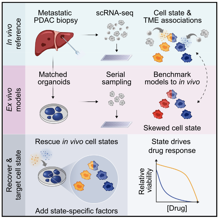
Prognostically relevant RNA expression states exist in pancreatic ductal adenocarcinoma (PDAC), but our understanding of their drivers, stability, and relationship to therapeutic response is limited. To examine these attributes systematically, we profiled metastatic biopsies and matched organoid models at single-cell resolution. In vivo, we identify a new intermediate PDAC transcriptional cell state and uncover distinct site- and state-specific tumor microenvironments (TMEs). Benchmarking models against this reference map, we reveal strong culture-specific biases in cancer cell transcriptional state representation driven by altered TME signals. We restore expression state heterogeneity by adding back in vivo-relevant factors and show plasticity in culture models. Further, we prove that non-genetic modulation of cell state can strongly influence drug responses, uncovering state-specific vulnerabilities. This work provides a broadly applicable framework for aligning cell states across in vivo and ex vivo settings, identifying drivers of transcriptional plasticity and manipulating cell state to target associated vulnerabilities.
Blood samples are frequently collected in human studies of the immune system but poorly represent tissue-resident immunity. Understanding the immunopathogenesis of tissue-restricted diseases, such as chronic hepatitis B, necessitates direct investigation of local immune responses. We developed a workflow that enables frequent, minimally invasive collection of liver fine-needle aspirates in multi-site international studies and centralized single-cell RNA sequencing data generation using the Seq-Well S3 picowell-based technology. All immunological cell types were captured, including liver macrophages, and showed distinct compartmentalization and transcriptional profiles, providing a systematic assessment of the capabilities and limitations of peripheral blood samples when investigating tissue-restricted diseases. The ability to electively sample the liver of chronic viral hepatitis patients and generate high-resolution data will enable multi-site clinical studies to power fundamental and therapeutic discovery.
Many patients infected with coronaviruses, such as SARS-CoV-2 and NL63 that use ACE2 receptors to infect cells, exhibit gastrointestinal symptoms and viral proteins are found in the human gastrointestinal tract, yet little is known about the inflammatory and pathological effects of coronavirus infection on the human intestine. Here, we used a human intestine-on-a-chip (Intestine Chip) microfluidic culture device lined by patient organoid-derived intestinal epithelium interfaced with human vascular endothelium to study host cellular and inflammatory responses to infection with NL63 coronavirus. These organoid-derived intestinal epithelial cells dramatically increased their ACE2 protein levels when cultured under flow in the presence of peristalsis-like mechanical deformations in the Intestine Chips compared to when cultured statically as organoids or in Transwell inserts. Infection of the intestinal epithelium with NL63 on-chip led to inflammation of the endothelium as demonstrated by loss of barrier function, increased cytokine production, and recruitment of circulating peripheral blood mononuclear cells (PBMCs). Treatment of NL63 infected chips with the approved protease inhibitor drug, nafamostat, inhibited viral entry and resulted in a reduction in both viral load and cytokine secretion, whereas remdesivir, one of the few drugs approved for COVID19 patients, was not found to be effective and it also was toxic to the endothelium. This model of intestinal infection was also used to test the effects of other drugs that have been proposed for potential repurposing against SARS-CoV-2. Taken together, these data suggest that the human Intestine Chip might be useful as a human preclinical model for studying coronavirus related pathology as well as for testing of potential anti-viral or anti-inflammatory therapeutics.
Existing preclinical methods for acquiring dissemination kinetics of rare circulating tumor cells (CTCs) en route to forming metastases have not been capable of providing a direct measure of CTC intravasation rate and subsequent half-life in the circulation. Here, we demonstrate an approach for measuring endogenous CTC kinetics by continuously exchanging CTC-containing blood over several hours between un-anesthetized, tumor-bearing mice and healthy, tumor-free counterparts. By tracking CTC transfer rates, we extrapolated half-life times in the circulation of between 40 and 260 s and intravasation rates between 60 and 107,000 CTCs/hour in mouse models of small-cell lung cancer (SCLC), pancreatic ductal adenocarcinoma (PDAC), and non-small cell lung cancer (NSCLC). Additionally, direct transfer of only 1−2% of daily-shed CTCs using our blood-exchange technique from late-stage, SCLC-bearing mice generated macrometastases in healthy recipient mice. We envision that our technique will help further elucidate the role of CTCs and the rate-limiting steps in metastasis.
A cell’s phenotype and function are influenced by dynamic interactions with its microenvironment. To examine cellular spatiotemporal activity, we developed SPACECAT—Spatially PhotoActivatable Color Encoded Cell Address Tags—to annotate, track, and isolate cells while preserving viability. In SPACECAT, samples are stained with photocaged fluorescent molecules, and cells are labeled by uncaging those molecules with user-patterned near-UV light. SPACECAT offers single-cell precision and temporal stability across diverse cell and tissue types. Illustratively, we target crypt-like regions in patient-derived intestinal organoids to enrich for stem-like and actively mitotic cells, matching literature expectations. Moreover, we apply SPACECAT to ex vivo tissue sections from four healthy organs and an autochthonous lung tumor model. Lastly, we provide a computational framework to identify spatially-biased transcriptome patterns and enriched phenotypes. This minimally perturbative and broadly applicable method links cellular spatiotemporal and/or behavioral phenotypes with diverse downstream assays, enabling insights into the connections between tissue microenvironments and (dys)function.
High-throughput single-cell RNA-sequencing (scRNA-seq) methodologies enable characterization of complex biological samples by increasing the number of cells that can be profiled contemporaneously. Nevertheless, these approaches recover less information per cell than low-throughput strategies. To accurately report the expression of key phenotypic features of cells, scRNA-seq platforms are needed that are both high fidelity and high throughput. To address this need, we created Seq-Well S3 (“Second-Strand Synthesis”), a massively parallel scRNA-seq protocol that uses a randomly primed second-strand synthesis to recover complementary DNA (cDNA) molecules that were successfully reverse transcribed but to which a second oligonucleotide handle, necessary for subsequent whole transcriptome amplification, was not appended due to inefficient template switching. Seq-Well S3 increased the efficiency of transcript capture and gene detection compared with that of previous iterations by up to 10- and 5-fold, respectively. We used Seq-Well S3 to chart the transcriptional landscape of five human inflammatory skin diseases, thus providing a resource for the further study of human skin inflammation.
Bulk transcriptomic studies have defined classical and basal-like gene expression subtypes in pancreatic ductal adenocarcinoma (PDAC) that correlate with survival and response to chemotherapy; however, the underlying mechanisms that govern these subtypes and their heterogeneity remain elusive. Here, we performed single-cell RNA-sequencing of 23 metastatic PDAC needle biopsies and matched organoid models to understand how tumor cell-intrinsic features and extrinsic factors in the tumor microenvironment (TME) shape PDAC cancer cell phenotypes. We identify a novel cancer cell state that co-expresses basal-like and classical signatures, demonstrates upregulation of developmental and KRAS-driven gene expression programs, and represents a transitional intermediate between the basal-like and classical poles. Further, we observe structure to the metastatic TME supporting a model whereby reciprocal intercellular signaling shapes the local microenvironment and influences cancer cell transcriptional subtypes. In organoid culture, we find that transcriptional phenotypes are plastic and strongly skew toward the classical expression state, irrespective of genotype. Moreover, we show that patient-relevant transcriptional heterogeneity can be rescued by supplementing organoid media with factors found in the TME in a subtype-specific manner. Collectively, our study demonstrates that distinct microenvironmental signals are critical regulators of clinically relevant PDAC transcriptional states and their plasticity, identifies the necessity for considering the TME in cancer modeling efforts, and provides a generalizable approach for delineating the cell-intrinsic versus -extrinsic factors that govern tumor cell phenotypes.
Barrier tissue epithelia play an essential role in maintaining organismal homeostasis, and changes in their cellular composition have been observed in multiple human diseases. Within the small intestinal epithelium, adult stem cells integrate diverse signals to regulate regeneration and differentiation, thereby establishing overall cellularity. Accordingly, directing stem cell differentiation could provide a tractable approach to alter the abundance or quality of specialized cells of the small intestinal epithelium, including the secretory Paneth, goblet, and enteroendocrine populations. Yet, to date, there has been a lack of suitable tools and rigorous approaches to identify biological targets and pharmacological agents that can modify epithelial composition to enable causal testing of disease-associated changes with novel therapeutic candidates. To empower the search for epithelia-modifying agents, we establish a first-of-its-kind high-throughput phenotypic organoid screen. We demonstrate the ability to screen thousands of samples and uncover biological targets and associated small molecule inhibitors which translate to in vivo. This approach is enabled by employing a functional, cell-type specific, scalable assay on an organoid model designed to represent the physiological cues of in vivo Paneth cell differentiation from adult intestinal stem cells. Further, we miniaturize and adapt the organoid culture system to enable automated plating and screening, thereby providing the ability to test thousands of samples. Strikingly, in our screen we identify inhibitors of the nuclear exporter Xpo1 modulate stem cell fate commitment by inducing a pan-epithelial stress response combined with an interruption of mitogen signaling in cycling intestinal progenitors, thereby significantly increasing the abundance of Paneth cells independent of known WNT and Notch differentiation cues. We extend our observation in vivo, demonstrating that oral administration of Xpo1 inhibitor KPT-330 at doses 1,000-fold lower than conventionally used in hematologic malignancies increases Paneth cell abundance. In total, we provide a framework to identify novel biological cues and therapeutic leads to rebalance intestinal stem cell differentiation and modulate epithelial tissue composition via high-throughput phenotypic screening in rationally-designed organoid model of differentiation.
The scale and capabilities of single-cell RNA-sequencing methods have expanded rapidly in recent years, enabling major dis- coveries and large-scale cell mapping efforts. However, these methods have not been systematically and comprehensively benchmarked. Here, we directly compare seven methods for single-cell and/or single-nucleus profiling—selecting representa- tive methods based on their usage and our expertise and resources to prepare libraries—including two low-throughput and five high-throughput methods. We tested the methods on three types of samples: cell lines, peripheral blood mononuclear cells and brain tissue, generating 36 libraries in six separate experiments in a single center. To directly compare the methods and avoid processing differences introduced by the existing pipelines, we developed scumi, a flexible computational pipeline that can be used with any single-cell RNA-sequencing method. We evaluated the methods for both basic performance, such as the structure and alignment of reads, sensitivity and extent of multiplets, and for their ability to recover known biological information in the samples.
Whether cultured in vitro or part of a complex tissue in vivo, a cell’s phenotype and function are significantly influenced by dynamic interactions with its microenvironment. To explicitly examine how a cell’s spatiotemporal activity impacts its behavior, we developed and validated a strategy termed SPACECAT – Spatially PhotoActivatable Color Encoded Cell Address Tags – to annotate, track, and isolate specific cells in a non-destructive, viability-preserving manner. In SPACECAT, a biological sample is immersed in a photocaged fluorescent molecule, and cells within a location of interest are labeled for further study by uncaging that molecule with user-patterned near-UV light. SPACECAT offers high spatial precision and temporal stability across diverse cell and tissue types, and is compatible with common downstream assays, including flow cytometry and single-cell RNA-Seq. Illustratively, we leveraged this approach in patient-derived intestinal organoids, a spatially complex system less amenable to genetic manipulations, to select for crypt-like regions enriched in stem-like and actively mitotic cells. Moreover, we demonstrate its applicability and utility on ex vivo tissue sections from four healthy organs and an autochthonous lung tumor model, uncovering spatially-biased gene expression patterns among immune cell subsets and identifying rare myeloid phenotypes enriched around tumor/healthy border regions. In sum, our method provides a minimally invasive and broadly applicable approach to link cellular spatiotemporal features and/or behavioral phenotypes with diverse downstream assays, enabling fundamental insights into the connections between tissue microenvironments and biological (dys)function.
By affinity capture and amplification of TCR transcripts from whole-transcriptome libraries, TCR CDR3 sequences can be recovered from 3′-barcoded scRNA-seq libraries (e.g. Seq-Well, Drop-seq, etc.). This method can be applied post-hoc, allowing for the capture of additional information from archived samples. The protocol can also be found here.
High-throughput 3′ single-cell RNA-sequencing (scRNA-seq) allows cost-effective, detailed characterization of individual immune cells from tissues. Current techniques, however, are limited in their ability to elucidate essential immune cell features, including variable sequences of T cell antigen receptors (TCRs) that confer antigen specificity. Here, we present a strategy that enables simultaneous analysis of TCR sequences and corresponding full transcriptomes from 3′-barcoded scRNA-seq samples. This approach is compatible with common 3′ scRNA-seq methods, and adaptable to processed samples post hoc. We applied the technique to identify transcriptional signatures associated with T cells sharing common TCRs from immunized mice and from patients with food allergy. We observed preferential phenotypes among subsets of expanded clonotypes, including type 2 helper CD4+ T cell (TH2) states associated with food allergy. These results demonstrate the utility of our method when studying diseases in which clonotype-driven responses are critical to understanding the underlying biology. The protocol can be found here.
To mark the 15th anniversary of Nature Methods, we asked scientists from across diverse fields of basic biology research for their views on the most exciting and essential methodological challenges that their communities are poised to tackle in the near future.
The development of high-throughput single-cell RNA-sequencing (scRNA-Seq) methodologies has empowered the characterization of complex biological samples by dramatically increasing the number of constituent cells that can be examined concurrently. Nevertheless, these approaches typically recover substantially less information per-cell as compared to lower-throughput microtiter plate-based strategies. To uncover critical phenotypic differences among cells and effectively link scRNA-Seq observations to legacy datasets, reliable detection of phenotype-defining transcripts – such as transcription factors, affinity receptors, and signaling molecules – by these methods is essential. Here, we describe a substantially improved massively-parallel scRNA-Seq protocol we term Seq-Well S^3 (“Second-Strand Synthesis”) that increases the efficiency of transcript capture and gene detection by up to 10- and 5-fold, respectively, relative to previous iterations, surpassing best-in-class commercial analogs. We first characterized the performance of Seq-Well S^3 in cell lines and PBMCs, and then examined five different inflammatory skin diseases, illustrative of distinct types of inflammation, to explore the breadth of potential immune and parenchymal cell states. Our work presents an essential methodological advance as well as a valuable resource for studying the cellular and molecular features that inform human skin inflammation.
Seq-Well is a low-cost picowell platform that can be used to simultaneously profile the transcriptomes of thousands of cells from diverse, low input clinical samples. In Seq-Well, uniquely barcoded mRNA capture beads and cells are co-confined in picowells that are sealed using a semipermeable membrane, enabling efficient cell lysis and mRNA capture. The beads are subsequently removed and processed in parallel for sequencing, with each transcript’s cell of origin determined via the unique barcodes. Due to its simplicity and portability, Seq-Well can be performed almost anywhere.
Circulating tumor cells (CTCs) play a fundamental role in cancer progression. However, in mice, limited blood volume and the rarity of CTCs in the bloodstream preclude longitudinal, in-depth studies of these cells using existing liquid biopsy techniques. Here, we present an optofluidic system that continuously collects fluorescently labeled CTCs from a genetically engineered mouse model (GEMM) for several hours per day over multiple days or weeks. The system is based on a microfluidic cell sorting chip connected serially to an unanesthetized mouse via an implanted arteriovenous shunt. Pneumatically controlled microfluidic valves capture CTCs as they flow through the device, and CTC-depleted blood is returned back to the mouse via the shunt. To demonstrate the utility of our system, we profile CTCs isolated longitudinally from animals over 4 days of treatment with the BET inhibitor JQ1 using single-cell RNA sequencing (scRNA-Seq) and show that our approach eliminates potential biases driven by intermouse heterogeneity that can occur when CTCs are collected across different mice. The CTC isolation and sorting technology presented here provides a research tool to help reveal details of how CTCs evolve over time, allowing studies to credential changes in CTCs as biomarkers of drug response and facilitating future studies to understand the role of CTCs in metastasis.
Mass and growth rate are highly integrative measures of cell physiology not discernable via genomic measurements. Here, we introduce a microfluidic platform enabling direct measurement of single-cell mass and growth rate upstream of highly multiplexed single-cell profiling such as single-cell RNA sequencing. We resolve transcriptional signatures associated with single-cell mass and growth rate in L1210 and FL5.12 cell lines and activated CD8+ T cells. Further, we demonstrate a framework using these linked measurements to characterize biophysical heterogeneity in a patient-derived glioblastoma cell line with and without drug treatment. Our results highlight the value of coupled phenotypic metrics in guiding single-cell genomics.
We introduce a microfluidic platform that enables single-cell mass and growth rate measurements upstream of single-cell RNA-sequencing (scRNA-seq) to generate paired single-cell biophysical and transcriptional data sets. Biophysical measurements are collected with a serial suspended microchannel resonator platform (sSMR) that utilizes automated fluidic state switching to load individual cells at fixed intervals, achieving a throughput of 120 cells per hour. Each single-cell is subsequently captured downstream for linked molecular analysis using an automated collection system. From linked measurements of a murine leukemia (L1210) and pro-B cell line (FL5.12), we identify gene expression signatures that correlate significantly with cell mass and growth rate. In particular, we find that both cell lines display a cell-cycle signature that correlates with cell mass, with early and late cell-cycle signatures significantly enriched amongst genes with negative and positive correlations with mass, respectively. FL5.12 cells also show a significant correlation between single-cell growth efficiency and a G1-S transition signature, providing additional transcriptional evidence for a phenomenon previously observed through biophysical measurements alone. Importantly, the throughput and speed of our platform allows for the characterization of phenotypes in dynamic cellular systems. As a proof-ofprinciple, we apply our system to characterize activated murine CD8+ T cells and uncover two unique features of CD8+ T cells as they become proliferative in response to activation: i) the level of coordination between cell cycle gene expression and cell mass increases, and ii) translation-related gene expression increases and shows a correlation with single-cell growth efficiency. Overall, our approach provides a new means of characterizing the transcriptional mechanisms of normal and dysfunctional cellular mass and growth rate regulation across a range of biological contexts
The recent advent of methods for high-throughput single-cell molecular profiling has catalyzed a growing sense in the scientific community that the time is ripe to complete the 150-year-old effort to identify all cell types in the human body. The Human Cell Atlas Project is an international collaborative effort that aims to define all human cell types in terms of distinctive molecular profiles (such as gene expression profiles) and to connect this information with classical cellular descriptions (such as location and morphology). An open comprehensive reference map of the molecular state of cells in healthy human tissues would propel the systematic study of physiological states, developmental trajectories, regulatory circuitry and interactions of cells, and also provide a framework for understanding cellular dysregulation in human disease. Here we describe the idea, its potential utility, early proofs-of-concept, and some design considerations for the Human Cell Atlas, including a commitment to open data, code, and community.
Single-cell RNA-seq could play a key role in personalized medicine by facilitating characterization of cells, pathways, and genes associated with human diseases such as cancer.
We develop single-cell transcriptomic approaches to comprehensively profile human tissues and model systems. Previously, we focused on establishing, validating, scaling, and simplifying single-cell RNA-seq, often through the development of microdevices, to enable genome-wide identification of the cell types/states contained within complex biological samples. More recently, we helped both enhance the detection of phenotype-defining transcripts using these methods and simplify their on-site processing for clinical applications. In parallel, we have also worked to democratize these techniques, providing open access to resources and protocols, training thousands locally and abroad, and establishing infrastructure and on-site collaborations spanning across 6 continents and 26+ countries.
As many factors define cellular phenotype and influence disease beyond mRNA, we develop complementary methods for co-assaying other cellular attributes to enrich our understanding of the drivers of cellular behaviors. Examples including the abundance of additional ‘-omes’, the sequence and amount of important transcripts, cellular history, biophysical properties, spatial position, and functional output. Recently, we have worked to: 1. detect pathogens in cells and potentially actionable associated host factors; 2. query for specific mutations to identify cancer cells; and, 3. extract T cell receptor sequences to examine clonality. We have also formulated computational methods to derive deeper insights from these data (e.g., to examine viral dynamic in infected cells, reproducible features hidden by inter-individual variability, multicellular immune dynamics, intercellular communication, or alteration in cellular ecosystems associated with pathology).
We explore how the extracellular milieu influences cellular decision-making. Here, we have employed controlled culture conditions with cells and organoids, chemical and genetic perturbations, and constant microfluidic perfusion. We also have leveraged natural microenvironmental variation within and across tissues via microdissection and by using photoactivatable probes that retain spatial information through dissociation. In each instance, we aim to understand the degree to which extracellular environments modulate, and can be used to rationally control, the responses of individual cells or the overall distribution thereof, with an eye toward engineering tissue responses.
We examine the impact of intercellular interactions on cellular function. We have used coculture, imaging and perturbation strategies, as well as matched computational methods, to reinforce findings from dissociated samples, validate inferred cell-cell communication in vivo (e.g., between sensory neurons and lymph node resident cells), and manipulate multicellular systems (e.g., organoids). We are currently working on building arrayed, synthetically-designed cellular ensembles to examine how ‘tissue’ structure impacts functional response. Our overall goal is to understand cellular co-dependencies that influence niche- and tissue-level response dynamics.
Recent advances in cellular profiling have demonstrated substantial heterogeneity in the behaviour of cells once deemed ‘identical’, challenging fundamental notions of cell ‘type’ and ‘state’. Not surprisingly, these findings have elicited substantial interest in deeply characterizing the diversity, interrelationships and plasticity among cellular phenotypes. To explore these questions, experimental platforms are needed that can extensively and controllably profile many individual cells. Here, microfluidic structures — whether valve-, droplet- or nanowell-based — have an important role because they can facilitate easy capture and processing of single cells and their components, reducing labour and costs relative to conventional plate-based methods while also improving consistency. In this article, we review the current state-of-the-art methodologies with respect to microfluidics for mammalian single-cell ‘omics’ and discuss challenges and future opportunities.
Single-cell RNA-seq can precisely resolve cellular states, but applying this method to low-input samples is challenging. Here, we present Seq-Well, a portable, low-cost platform for massively parallel single-cell RNA-seq. Barcoded mRNA capture beads and single cells are sealed in an array of subnanoliter wells using a semipermeable membrane, enabling efficient cell lysis and transcript capture. We use Seq-Well to profile thousands of primary human macrophages exposed to Mycobacterium tuberculosis.
We lack effective treatments and preventions for many of the most challenging infectious diseases, many of which disproportionately impact those in low- and middle-income countries or traditionally marginalized communities.
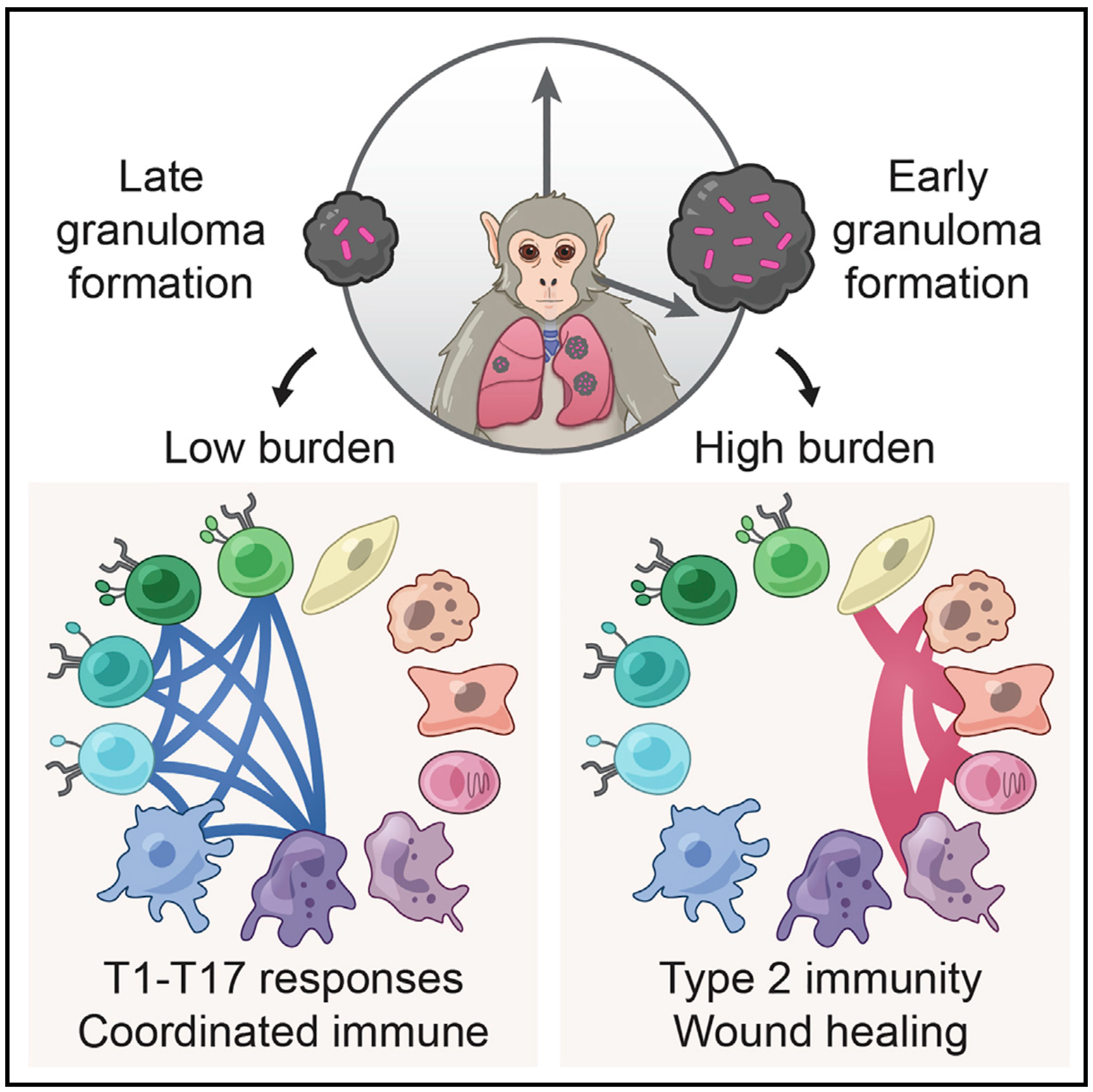
To help address this, we have established and enabled multi-group, multi-country partnerships to deploy and adapt cutting-edge genomic tools. By examining how cells dynamically alter their states, individually and collectively, during disease and/or its resolution in acute and chronic infections—e.g., tuberculosis, HIV/SHIV, hepatitis, malaria, leprosy, flu, SARS-CoV-2, and ebola—we have uncovered cellular and molecular features of pathogen control or pathology to potentiate or counteract, respectively. Illustratively, in tuberculosis, we identified a functional role for cytotoxic CD8 and hybrid type1-type17 T cells in control of infection in the lung and links between mast, plasma, and endothelial cell abundance (type-2 immune responses) and bacterial burden. We have also built methods for examining pathogens within individual host cells to define their dynamic interdependence and identify potentially restrictive host factors.
We are currently working to identify the drivers of common host responses to distinct perturbations and their targetability, as well as the impact of different interventions (e.g., vaccines).
Immune responses play a critical role in preventing tumorigenesis. Sometimes, however, they are ineffectual and can even drive/support malignancy.
We have examined how cancer cells alter and are influenced by their tumor microenvironments (TMEs), and the impact this has on therapeutic responses. Illustratively, in Pancreatic Ductal Adenocarcinoma (PDAC), by profiling liver metastases and matched organoid models, we showed: 1. associations between TME and malignant cell state composition; 2. that autocrine and paracrine signaling can drive malignant cell state transitions, even in an isogenic background, altering the efficacy of frontline chemotherapies; and, 3. that microenvironmental manipulations can be used to control malignant state, and thereby drug responses, rationally, and to improve model fidelity for screening potential therapies. This and related work highlight the potential utility of modulating indirect target cells (T cells in the PDAC TME or basal cells in allergic inflammation) to enhance cures and preventions.
We are now systematically expanding this work to define how additional environmental and cell-intrinsic factors influence malignant cell state plasticity in PDAC and other cancers toward enhancing treatments.
We are exposed to a constant flux of external biochemical and physical stimuli as we age. Despite variability in our overall experiences and exact constitutions, our individual tissues typically manage to maintain functionality, though each can differ in its resilience to distinct stressors.
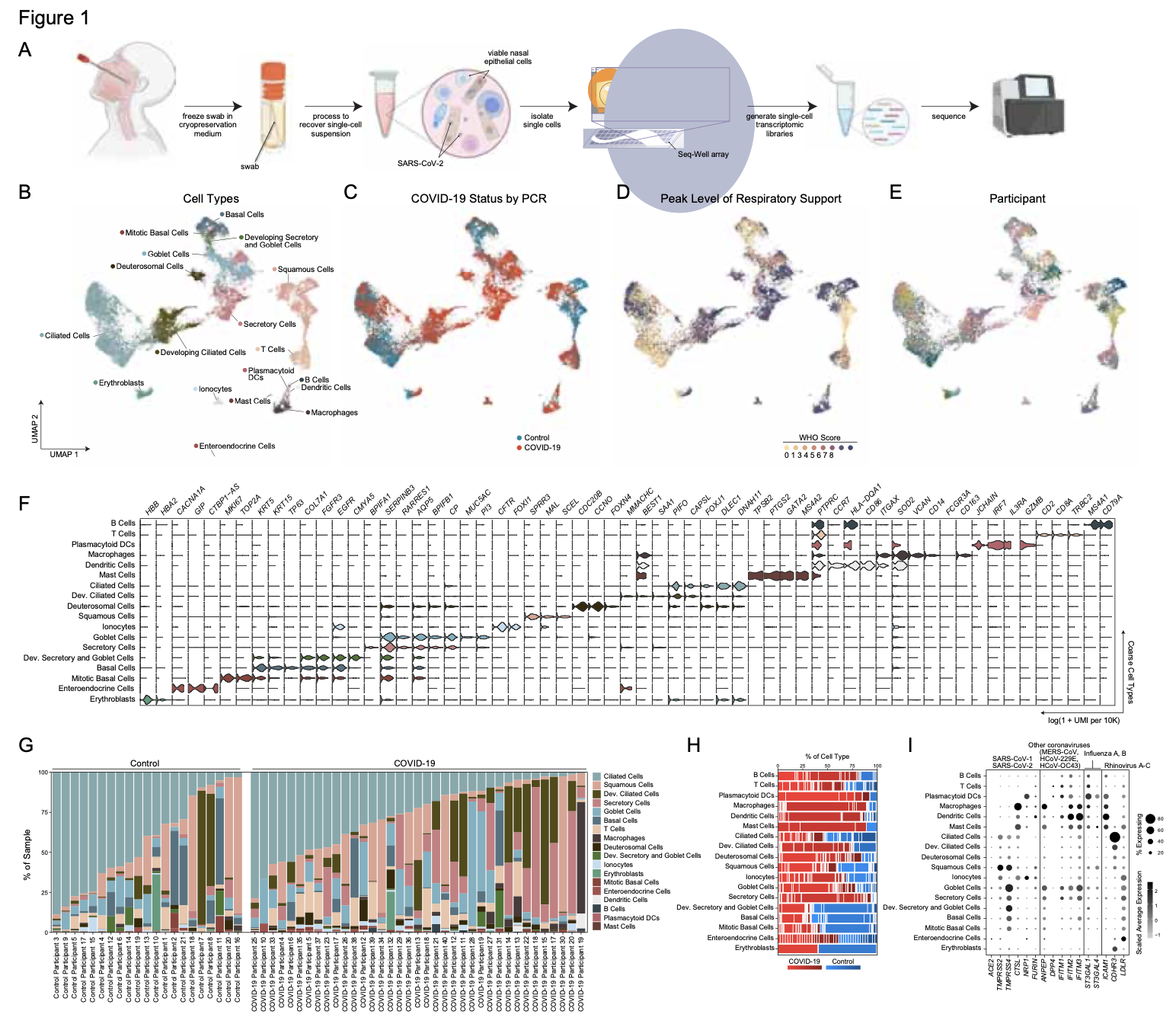
We have characterized how differences in cellular composition and communication impact tissue fitness and have identified responses and subsequent adaptations that drive chronic dysfunction. For example, although aberrant immune activity can precipitate allergic inflammatory diseases, therapies targeting immune cells and signaling are only successful in some, suggesting chronicity may involve alternative mechanisms. Previously, we helped demonstrate that dysregulated type-2 immune signaling, driven by environmental allergens, can impact tissue health in the upper airway through generating dysfunctional basal epithelial stem cells. These stem cells can then contribute to persistence by serving as repositories for allergic inflammatory memories, altering the integrity and functional output of the nasal epithelium. Our work, with that of others, suggests generalizable principles for cellular memory, and informs where and how tissues should be targeted to support health or restore function. We have since further investigated how tissue-resident cellular subsets participate in, and are shaped by, environmental exposures at barrier tissues and the functional consequences of these experiences.
We are now working to develop a more holistic appreciation for how different intra- and extracellular factors (e.g., genetics and integrated exposure history, respectively) influence barrier tissue function.
We present a scalable, integrated strategy for coupled protein and RNA detection from single cells. Our approach leverages the DNA polymerase activity of reverse transcriptase to simultaneously perform proximity extension assays and complementary DNA synthesis in the same reaction. Using the Fluidigm C1TM system, we profile the transcriptomic and proteomic response of a human breast adenocarcinoma cell line to a chemical perturbation, benchmarking against in situ hybridizations and immunofluorescence staining, as well as recombinant proteins, ERCC Spike-Ins, and population lysate dilutions. Through supervised and unsupervised analyses, we demonstrate synergies enabled by simultaneous measurement of single-cell protein and RNA abundances. Collectively, our generalizable approach highlights the potential for molecular metadata to inform highly-multiplexed single-cell analyses.
The orchestrated action of genes controls complex biological phenotypes, yet the systematic discovery of gene and drug combinations that modulate these phenotypes in human cells is labor intensive and challenging to scale. Here, we created a platform for the massively parallel screening of barcoded combinatorial gene perturbations in human cells and translated these hits into effective drug combinations. This technology leverages the simplicity of the CRISPR-Cas9 system for multiplexed targeting of specific genomic loci and the versatility of combinatorial genetics en masse (CombiGEM) to rapidly assemble barcoded combinatorial genetic libraries that can be tracked with high-throughput sequencing. We applied CombiGEM-CRISPR to create a library of 23,409 barcoded dual guide-RNA (gRNA) combinations and then perform a high-throughput pooled screen to identify gene pairs that inhibited ovarian cancer cell growth when they were targeted. We validated the growth-inhibiting effects of specific gene sets, including epigenetic regulators KDM4C/BRD4 and KDM6B/BRD4, via individual assays with CRISPR-Cas–based knockouts and RNA-interference–based knockdowns. We also tested small-molecule drug pairs directed against our pairwise hits and showed that they exerted synergistic antiproliferative effects against ovarian cancer cells. We envision that the CombiGEM-CRISPR platform will be applicable to a broad range of biological settings and will accelerate the systematic identification of genetic combinations and their translation into novel drug combinations that modulate complex human disease phenotypes.
We introduce a microfluidic platform that enables off-chip single-cell RNA-seq after multi-generational lineage tracking under controlled culture conditions. We use this platform to generate whole-transcriptome profiles of primary, activated murine CD8+ T-cell and lymphocytic leukemia cell line lineages. Here we report that both cell types have greater intra- than inter-lineage transcriptional similarity. For CD8+ T-cells, genes with functional annotation relating to lymphocyte differentiation and function—including Granzyme B—are enriched among the genes that demonstrate greater intra-lineage expression level similarity. Analysis of gene expression covariance with matched measurements of time since division reveals cell type-specific transcriptional signatures that correspond with cell cycle progression. We believe that the ability to directly measure the effects of lineage and cell cycle-dependent transcriptional profiles of single cells will be broadly useful to fields where heterogeneous populations of cells display distinct clonal trajectories, including immunology, cancer, and developmental biology.
Encounters between immune cells and invading bacteria ultimately determine the course of infection. These interactions are usually measured in populations of cells, masking cell-to-cell variation that may be important for infection outcome. To characterize the gene expression variation that underlies distinct infection outcomes and monitor infection phenotypes, we developed an experimental system that combines single-cell RNA-seq with fluorescent markers. Probing the responses of individual macrophages to invading Salmonella, we find that variation between individual infected host cells is determined by the heterogeneous activity of bacterial factors in individual infecting bacteria. We illustrate how variable PhoPQ activity in the population of invading bacteria drives variable host type I IFN responses by modifying LPS in a subset of bacteria. This work demonstrates a causative link between host and bacterial variability, with cell-to-cell variation between different bacteria being sufficient to drive radically different host immune responses. This co-variation has implications for host-pathogen dynamics in vivo.
An innovative, microwell-based platform for single-cell RNA sequencing (RNA-seq) combines cost efficiency, scalability and parallelizability, and will enable many new avenues of biological inquiry.
Cells, the basic units of biological structure and function, vary broadly in type and state. Single- cell genomics can characterize cell identity and function, but limitations of ease and scale have prevented its broad application. Here we describe Drop-seq, a strategy for quickly profiling thousands of individual cells by separating them into nanoliter-sized aqueous droplets, associating a different barcode with each cell’s RNAs, and sequencing them all together. Drop-seq analyzes mRNA transcripts from thousands of individual cells simultaneously while remembering transcripts’ cell of origin. We analyzed transcriptomes from 44,808 mouse retinal cells and identified 39 transcriptionally distinct cell populations, creating a molecular atlas of gene expression for known retinal cell classes and novel candidate cell subtypes. Drop-seq will accelerate biological discovery by enabling routine transcriptional profiling at single- cell resolution.
For the past several decades, due to technical limitations, the field of transcriptomics has focused on population-level measurements that can mask significant differences between individual cells. With the advent of single-cell RNA-Seq, it is now possible to profile the responses of individual cells at unprecedented depth and thereby uncover, transcriptome-wide, the heterogeneity that exists within these populations. This unit describes a method that merges several important technologies to produce, in highthroughput, single-cell RNA-Seq libraries. Complementary DNA (cDNA) is made from full-length mRNA transcripts using a reverse transcriptase that has terminal transferase activity. This,when combinedwith a second “template-switch” primer, allows for cDNAs to be constructed that have two universal priming sequences. Following preamplification from these common sequences, Nextera XT is used to prepare a pool of 96 uniquely indexed samples ready for Illumina sequencing.
The mammalian immune system is tasked with protecting the host against a broad range of threats. Understanding how immune populations leverage cellular diversity to achieve this breadth and flexibility, particularly during dynamic processes such as differentiation and antigenic response, is a core challenge that is well suited for single cell analysis. Recent years have witnessed transformative and intersecting advances in nanofabrication and genomics that enable deep profiling of individual cells, affording exciting opportunities to study heterogeneity in the immune response at an unprecedented scope. In light of these advances, here we review recent work exploring how immune populations generate and leverage cellular heterogeneity at multiple molecular and phenotypic levels. Additionally, we highlight opportunities for single cell technologies to shed light on the causes and consequences of heterogeneity in the immune system.
Developing a detailed understanding of enzyme function in the context of an intracellular signal transduction pathway requires minimally invasive methods for probing enzyme activity in situ. Here, we describe a new method for monitoring enzyme activity in living cells by sandwiching live cells between two vertical silicon nanowire (NW) arrays. Specifically, we use the first NW array to immobilize the cells and then present enzymatic substrates intracellularly via the second NW array by utilizing the NWs’ ability to penetrate cellular membranes without affecting cells’ viability or function. This strategy, when coupled with fluorescence microscopy and mass spectrometry, enables intracellular examination of protease, phosphatase, and protein kinase activities, demonstrating the assay’s potential in uncovering the physiological roles of various enzymes.
A circuit level understanding of immune cells and hematological cancers has been severely impeded by a lack of techniques that enable intracellular perturbation without significantly altering cell viability and function. Here, we demonstrate that vertical silicon nanowires (NWs) enable gene-specific manipulation of diverse murine and human immune cells with negligible toxicity. To illustrate the power of the technique, we then apply NW-mediated gene silencing to investigate the role of the Wnt signaling pathway in chronic lymphocytic leukemia (CLL). Remarkably, CLL-B cells from different patients exhibit tremendous heterogeneity in their response to the knockdown of a single gene, LEF1. This functional heterogeneity defines three distinct patient groups not discernible by conventional CLL cytogenetic markers and provides a prognostic indicator for patients’ time to first therapy. Analyses of gene expression signatures associated with these functional patient subgroups reveal unique insights into the underlying molecular basis for disease heterogeneity. Overall, our findings suggest a functional classification that can potentially guide the selection of patient-specific therapies in CLL and highlight the opportunities for nanotechnology to drive biological inquiry.
Deciphering the neuronal code—the rules by which neuronal circuits store and process information—is a major scientific challenge. Currently, these efforts are impeded by a lack of experimental tools that are sensitive enough to quantify the strength of individual synaptic connections and also scalable enough to simultaneously measure and control a large number of mammalian neurons with single-cell resolution. Here, we report a scalable intracellular electrode platform based on vertical nanowires that allows parallel electrical interfacing to multiple mammalian neurons. Specifically, we show that our vertical nanowire electrode arrays can intracellularly record and stimulate neuronal activity in dissociated cultures of rat cortical neurons and can also be used to map multiple individual synaptic connections. The scalability of this platform, combined with its compatibility with silicon nanofabrication techniques, provides a clear path towards simultaneous, highfidelity interfacing with hundreds of individual neurons.
A generalized platform for introducing a diverse range of biomolecules into living cells in high-throughput could transform how complex cellular processes are probed and analyzed. Here, we demonstrate spatially localized, efficient, and universal delivery of biomolecules into immortalized and primary mammalian cells using surface-modified vertical silicon nanowires. The method relies on the ability of the silicon nanowires to penetrate a cell’s membrane and subsequently release surface-bound molecules directly into the cell’s cytosol, thus allowing highly efficient delivery of biomolecules without chemical modification or viral packaging. This modality enables one to assess the phenotypic consequences of introducing a broad range of biological effectors (DNAs, RNAs, peptides, proteins, and small molecules) into almost any cell type. We show that this platform can be used to guide neuronal progenitor growth with small molecules, knock down transcript levels by delivering siRNAs, inhibit apoptosis using peptides, and introduce targeted proteins to specific organelles. We further demonstrate codelivery of siRNAs and proteins on a single substrate in a microarray format, highlighting this technology’s potential as a robust, monolithic platform for high-throughput, miniaturized bioassays.
Abstract
Electrostatic force microscopy shows that the electric field gradients above pentacene monolayer islands on 2-nm SiO2/Si substrates, in a dark, dry nitrogen environment, display a wide distribution of signs and magnitude that is dependent on sample history. Under 12 mW/cm2 green (532 nm) illumination, pentacene islands accumulate positive charge because of photoexcited electron transfer across the oxide to the Si substrate. At a strong illumination of 60 mW/cm2, pentacene islands reform into small spherical particles, apparently because the positive charge Coulomb repulsion energy becomes comparable to the cohesive energy of the pentacene monolayer.