Genome-wide association studies (GWAS) have revealed risk alleles for ulcerative colitis (UC). To understand their cell type specificities and pathways of action, we generate an atlas of 366,650 cells from the colon mucosa of 18 UC patients and 12 healthy individuals, revealing 51 epithelial, stromal, and immune cell subsets, including BEST4+ enterocytes, microfold-like cells, and IL13RA2+IL11+ inflammatory fibroblasts, which we associate with resistance to anti-TNF treatment. Inflammatory fibroblasts, inflammatory monocytes, microfold-like cells, and T cells that co-express CD8 and IL-17 expand with disease, forming intercellular interaction hubs. Many UC risk genes are cell type specific and co-regulated within relatively few gene modules, suggesting convergence onto limited sets of cell types and pathways. Using this observation, we nominate and infer functions for specific risk genes across GWAS loci. Our work provides a framework for interrogating complex human diseases and mapping risk variants to cell types and pathways.

The development of high-throughput single-cell RNA-sequencing (scRNA-Seq) methodologies has empowered the characterization of complex biological samples by dramatically increasing the number of constituent cells that can be examined concurrently. Nevertheless, these approaches typically recover substantially less information per-cell as compared to lower-throughput microtiter plate-based strategies. To uncover critical phenotypic differences among cells and effectively link scRNA-Seq observations to legacy datasets, reliable detection of phenotype-defining transcripts – such as transcription factors, affinity receptors, and signaling molecules – by these methods is essential. Here, we describe a substantially improved massively-parallel scRNA-Seq protocol we term Seq-Well S^3 (“Second-Strand Synthesis”) that increases the efficiency of transcript capture and gene detection by up to 10- and 5-fold, respectively, relative to previous iterations, surpassing best-in-class commercial analogs. We first characterized the performance of Seq-Well S^3 in cell lines and PBMCs, and then examined five different inflammatory skin diseases, illustrative of distinct types of inflammation, to explore the breadth of potential immune and parenchymal cell states. Our work presents an essential methodological advance as well as a valuable resource for studying the cellular and molecular features that inform human skin inflammation.

Tuberculosis is the leading cause of death by an infectious disease worldwide1. However, the involvement of innate lymphoid cells (ILCs) in immune responses to infection with Mycobacterium tuberculosis (Mtb) is unknown. Here we show that circulating subsets of ILCs are depleted from the blood of participants with pulmonary tuberculosis and restored upon treatment. Tuberculosis increased accumulation of ILC subsets in the human lung, coinciding with a robust transcriptional response to infection, including a role in orchestrating the recruitment of immune subsets. Using mouse models, we show that group 3 ILCs (ILC3s) accumulated rapidly in Mtb-infected lungs and coincided with the accumulation of alveolar macrophages. Notably, mice that lacked ILC3s exhibited a reduction in the accumulation of early alveolar macrophages and decreased Mtb control. We show that the C-X-C motif chemokine receptor 5 (CXCR5)–C-X-C motif chemokine ligand 13 (CXCL13) axis is involved in Mtb control, as infection upregulates CXCR5 on circulating ILC3s and increases plasma levels of its ligand, CXCL13, in humans. Moreover, interleukin-23-dependent expansion of ILC3s in mice and production of interleukin-17 and interleukin-22 were found to be critical inducers of lung CXCL13, early innate immunity and the formation of protective lymphoid follicles within granulomas. Thus, we demonstrate an early protective role for ILC3s in immunity to Mtb infection.

Cellular immunity is critical for controlling intracellular pathogens, but the dynamics and cooperativity of the evolving host response to infection are not well defined. Here, we apply single-cell RNA-sequencing to longitudinally profile pre- and immediately post-HIV infection peripheral immune responses of multiple cell types in four untreated individuals. Onset of viremia induces a strong transcriptional interferon response integrated across most cell types, with subsequent pro-inflammatory T cell differentiation, monocyte MHC-II upregulation, and cytolytic killing. With longitudinal sampling, we nominate key intra- and extracellular drivers that induce these programs, and assign their multi-cellular targets, temporal ordering, and duration in acute infection. Two individuals studied developed spontaneous viral control, associated with initial elevated frequencies of proliferating cytotoxic cells, inclusive of a previously unappreciated proliferating natural killer (NK) cell subset. Our study presents a unified framework for characterizing immune evolution during a persistent human viral infection at single-cell resolution, and highlights programs that may drive response coordination and influence clinical trajectory.

Sustained viremia after acute HIV infection is associated with profound CD4+ T cell loss and exhaustion of HIV-specific CD8+ T cell responses. To determine the impact of combination antiretroviral therapy (cART) on these processes, we examined the evolution of immune responses in acutely infected individuals initiating treatment before peak viremia. Immediate treatment of Fiebig stages I and II infection led to a rapid decline in viral load and diminished magnitude of HIV-specific (tetramer+) CD8+ T cell responses compared to untreated donors. There was a strong positive correlation between cumulative viral antigen exposure before full cART-induced suppression and immune responses measured by MHC class I tetramers, IFN-γ ELISPOT, and CD8+ T cell activation. HIV-specific CD8+ T responses of early treated individuals were characterized by increased CD127 and BCL-2 expression, greater in vitro IFN-γ secretion, and enhanced differentiation into effector memory (Tem) cells. Transcriptional analysis of tetramer+ CD8+ T cells from treated persons revealed reduced expression of genes associated with activation and apoptosis, with concurrent up-regulation of prosurvival genes including BCL-2AXL, and SRC. Early treatment also resulted in robust HIV-specific CD4+ T cell responses compared to untreated HIV-infected individuals. Our data show that limiting acute viremia results in enhanced functionality of HIV-specific CD4+ and CD8+ T cells, preserving key antiviral properties of these cells.

The extraordinary activity of high-dose cyclophosphamide against some high-grade lymphomas was described nearly 60 years ago. Here we address mechanisms that mediate cyclophosphamide activity in bona fide human double-hit lymphoma. We show that antibody resistance within the bone marrow (BM) is not present upon early engraftment but develops during lymphoma progression. This resistance required a high tumor: macrophage ratio, was recapitulated in spleen by partial macrophage depletion and was overcome by multiple, highdose alkylating agents. Cyclophosphamide induced ER-stress in BM-resident lymphoma cells in vivo that resulted in ATF4-mediated paracrine secretion of VEGF-A, massive macrophage infiltration and clearance of alemtuzumab-opsonized cells. BM macrophages isolated after cyclophosphamide treatment had increased phagocytic capacity that was reversed by VEGF-A blockade or SYK inhibition. Single-cell RNA sequencing of these macrophages identified a “super-phagocytic” subset that expressed CD36/FcgRIV. Together, these findings define a novel mechanism through which high-dose alkylating agents promote macrophage-dependent lymphoma clearance.

Mice engrafted with components of a human immune system have become widely-used models for studying aspects of human immunity and disease. However, a defined methodology to objectively measure and compare the quality of the human immune response in different models is lacking. Here, by taking advantage of the highly immunogenic live-attenuated yellow fever virus vaccine YFV-17D, we provide an in-depth comparison of immune responses in human vaccinees, conventional humanized mice, and second generation humanized mice. We demonstrate that selective expansion of human myeloid and natural killer cells promotes transcriptomic responses akin to those of human vaccinees. These enhanced transcriptomic profiles correlate with the development of an antigen-specific cellular and humoral response to YFV-17D. Altogether, our approach provides a robust scoring of the quality of the human immune response in humanized mice and highlights a rational path towards developing better pre-clinical models for studying the human immune response and disease.

In the small intestine, a niche of accessory cell types supports the generation of mature epithelial cell types from intestinal stem cells (ISCs). It is unclear, however, if and how immune cells in the niche affect ISC fate or the balance between self-renewal and differentiation. Here, we use single-cell RNA sequencing (scRNA-seq) to identify MHC class II (MHCII) machinery enrichment in two subsets of Lgr5 + ISCs. We show that MHCII +Lgr5 + ISCs are non-conventional antigen-presenting cells in co-cultures with CD4 + T helper (Th) cells. Stimulation of intestinal organoids with key Th cytokines affects Lgr5 + ISC renewal and differentiation in opposing ways: pro-inflammatory signals promote differentiation, while regulatory cells and cytokines reduce it. In vivo genetic perturbation of Th cells or MHCII expression on Lgr5 + ISCs impacts epithelial cell differentiation and IEC fate during infection. These interactions between Th cells and Lgr5 + ISCs, thus, orchestrate tissue-wide responses to external signals.

Genome-wide association studies (GWAS) have revealed risk alleles for ulcerative colitis (UC), but their cell type and pathway specificities are often unknown. Here, we generate an atlas of 115,517 cells from the colon mucosa of seven UC patients and ten healthy individuals, revealing 51 epithelial, stromal, and immune cell subsets, including a subset of BEST4+ enterocytes, which may sense and respond to pH, and IL13RA2+IL-11+ inflammatory fibroblasts, which we associate with resistance to anti-TNF therapy. Inflammatory fibroblasts, inflammatory monocytes, microfold-like cells, and CD8+IL-17+ T cells expand during disease, and form intercellular interaction hubs that mediate cross-talk between diverse cellular lineages. We identify hundreds of putative autocrine and paracrine cell-cell interactions that may explain the migration, expansion, or inhibition of cell types with disease. Surprisingly, UC risk genes are often cell type specific and co-regulated in relatively few gene modules, suggesting convergence onto limited sets of cell types and pathways. Using this observation, we nominate and infer putative functions for UC risk genes across all GWAS loci. Our atlas thus provides a framework for interrogating complex human diseases and mapping risk variants onto their cell types and pathways of activity.

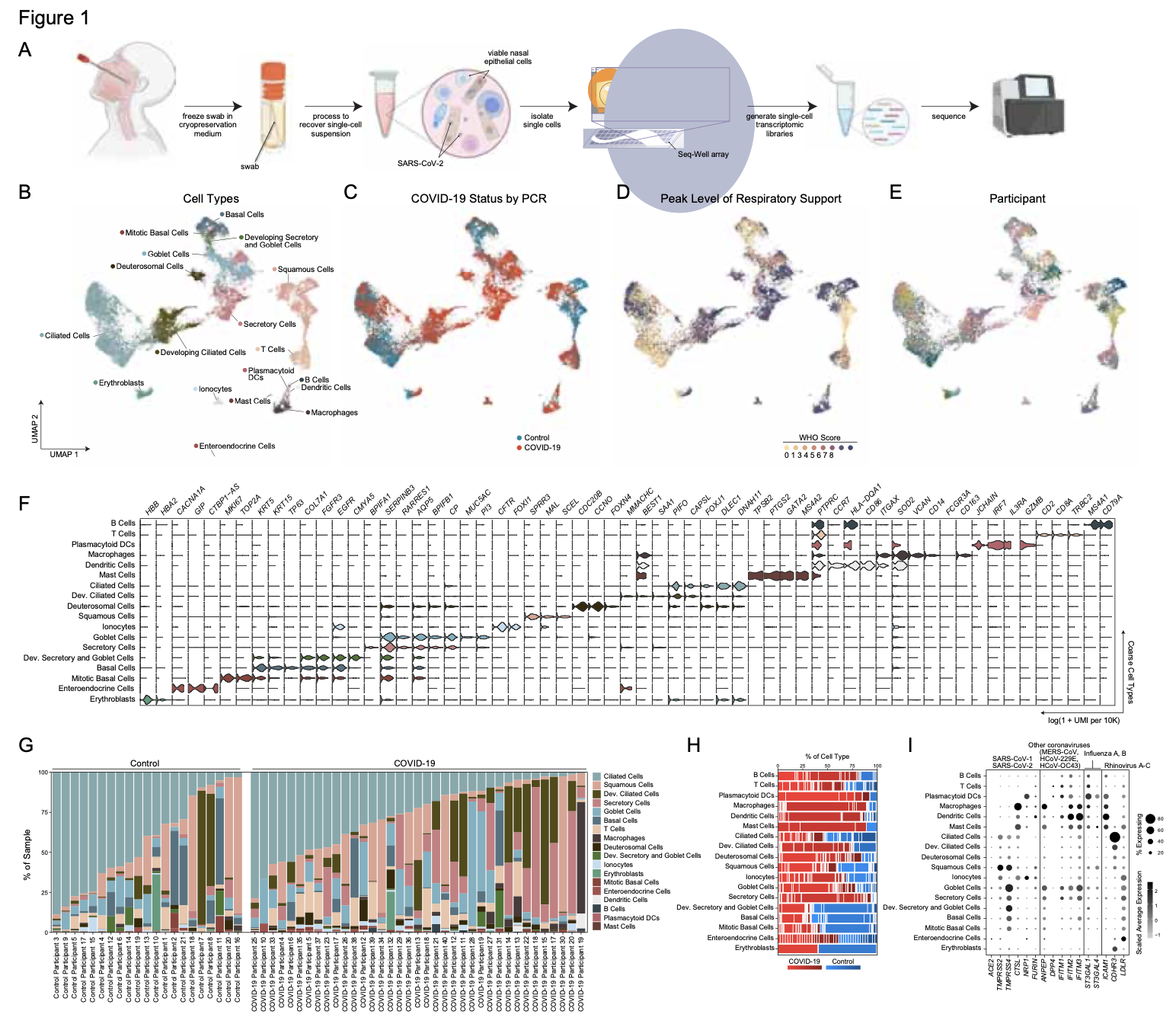
Barrier tissue dysfunction is a fundamental feature of chronic human inflammatory diseases1. Specialized subsets of epithelial cells—including secretory and ciliated cells—differentiate from basal stem cells to collectively protect the upper airway2,3,4. Allergic inflammation can develop from persistent activation5 of type 2 immunity6 in the upper airway, resulting in chronic rhinosinusitis, which ranges in severity from rhinitis to severe nasal polyps7. Basal cell hyperplasia is a hallmark of severe disease7,8,9, but it is not known how these progenitor cells2,10,11contribute to clinical presentation and barrier tissue dysfunction in humans. Here we profile primary human surgical chronic rhinosinusitis samples (18,036 cells, n = 12) that span the disease spectrum using Seq-Well for massively parallel single-cell RNA sequencing12, report transcriptomes for human respiratory epithelial, immune and stromal cell types and subsets from a type 2 inflammatory disease, and map key mediators. By comparison with nasal scrapings (18,704 cells, n = 9), we define signatures of core, healthy, inflamed and polyp secretory cells. We reveal marked differences between the epithelial compartments of the non-polyp and polyp cellular ecosystems, identifying and validating a global reduction in cellular diversity of polyps characterized by basal cell hyperplasia, concomitant decreases in glandular cells, and phenotypic shifts in secretory cell antimicrobial expression. We detect an aberrant basal progenitor differentiation trajectory in polyps, and propose cell-intrinsic13, epigenetic14,15 and extrinsic factors11,16,17 that lock polyp basal cells into this uncommitted state. Finally, we functionally demonstrate that ex vivo cultured basal cells retain intrinsic memory of IL-4/IL-13 exposure, and test the potential for clinical blockade of the IL-4 receptor α-subunit to modify basal and secretory cell states in vivo. Overall, we find that reduced epithelial diversity stemming from functional shifts in basal cells is a key characteristic of type 2 immune-mediated barrier tissue dysfunction. Our results demonstrate that epithelial stem cells may contribute to the persistence of human disease by serving as repositories for allergic memories.

Background

Human immunity relies on the coordinated responses of many cellular subsets and functional states. Inter-individual variations in cellular composition and communication could thus potentially alter host protection. Here, we explore this hypothesis by applying single-cell RNA-sequencing to examine viral responses among the dendritic cells (DCs) of three elite controllers (ECs) of HIV-1 infection.

Results

To overcome the potentially confounding effects of donor-to-donor variability, we present a generally applicable computational framework for identifying reproducible patterns in gene expression across donors who share a unifying classification. Applying it, we discover a highly functional antiviral DC state in ECs whose fractional abundance after in vitro exposure to HIV-1 correlates with higher CD4+ T cell counts and lower HIV-1 viral loads, and that effectively primes polyfunctional T cell responses in vitro. By integrating information from existing genomic databases into our reproducibility-based analysis, we identify and validate select immunomodulators that increase the fractional abundance of this state in primary peripheral blood mononuclear cells from healthy individuals in vitro.

Conclusions

Overall, our results demonstrate how single-cell approaches can reveal previously unappreciated, yet important, immune behaviors and empower rational frameworks for modulating systems-level immune responses that may prove therapeutically and prophylactically useful.


Complete information about the scRAD R package is available on the Shalek Lab Resources page.

B cells constitute an essential line of defense from pathogenic infections through the generation of class-switched antibody-secreting cells (ASCs) in germinal centers. Although this process is known to be regulated by follicular helper T (TfH) cells, the mechanism by which B cells initially seed germinal center reactions remains elusive. We found that NKT cells, a population of innate-like T lymphocytes, are critical for the induction of B cell immunity upon viral infection. The positioning of NKT cells at the interfollicular areas of lymph nodes facilitates both their direct priming by resident macrophages and the localized delivery of innate signals to antigen-experienced B cells. Indeed, NKT cells secrete an early wave of IL-4 and constitute up to 70% of the total IL-4-producing cells during the initial stages of infection. Importantly, the requirement of this innate immunity arm appears to be evolutionarily conserved because early NKT and IL-4 gene signatures also positively correlate with the levels of neutralizing antibodies in Zika-virus-infected macaques. In conclusion, our data support a model wherein a pre-TfH wave of IL-4 secreted by interfollicular NKT cells triggers the seeding of germinal center cells and serves as an innate link between viral infection and B cell immunity.

The recent advent of methods for high-throughput single-cell molecular profiling has catalyzed a growing sense in the scientific community that the time is ripe to complete the 150-year-old effort to identify all cell types in the human body. The Human Cell Atlas Project is an international collaborative effort that aims to define all human cell types in terms of distinctive molecular profiles (such as gene expression profiles) and to connect this information with classical cellular descriptions (such as location and morphology). An open comprehensive reference map of the molecular state of cells in healthy human tissues would propel the systematic study of physiological states, developmental trajectories, regulatory circuitry and interactions of cells, and also provide a framework for understanding cellular dysregulation in human disease. Here we describe the idea, its potential utility, early proofs-of-concept, and some design considerations for the Human Cell Atlas, including a commitment to open data, code, and community.

Tissue barrier dysfunction is a poorly defined feature hypothesized to drive chronic human inflammatory disease. The epithelium of the upper respiratory tract represents one such barrier, responsible for separating inhaled agents, such as pathogens and allergens, from the underlying submucosa. Specialized epithelial subsets-including secretory, glandular, and ciliated cells-differentiate from basal progenitors to collectively realize this role. Allergic inflammation in the upper airway barrier can develop from persistent activation of Type 2 immunity (T2I), resulting in the disease spectrum known as chronic rhinosinusitis (CRS), ranging from rhinitis to severe nasal polyps. Whether recently identified epithelial progenitor subsets, and their differentiation trajectory, contribute to the clinical presentation and barrier dysfunction in T2I-mediated disease in humans remains unexplored. Profiling twelve primary human CRS samples spanning the range of clinical severity with the Seq-Well platform for massively-parallel single-cell RNA-sequencing (scRNA-seq), we report the first single-cell transcriptomes for human respiratory epithelial cell subsets, immune cells, and parenchymal cells (18,036 total cells) from a T2I inflammatory disease, and map key mediators. We find striking differences between non-polyp and polyp tissues within the epithelial compartments of human T2I cellular ecosystems. More specifically, across 10,383 epithelial cells, we identify a global reduction in epithelial diversity in polyps characterized by basal cell hyperplasia, a concomitant decrease in glandular and ciliated cells, and phenotypic shifts in secretory cell function. We validate these findings through flow cytometry, histology, and bulk tissue RNA-seq of an independent cohort. Furthermore, we detect an aberrant basal progenitor differentiation trajectory in polyps, and uncover cell-intrinsic and extrinsic factors that may lock polyp basal cells into an uncommitted state. Overall, our data define severe T2I barrier dysfunction as a reduction in epithelial diversity, characterized by profound functional shifts stemming from basal cell defects, and nominate a cellular mechanism for the persistence and chronicity of severe human respiratory disease.

Click here to read the pre-publication manuscript.

Single-cell RNA-seq could play a key role in personalized medicine by facilitating characterization of cells, pathways, and genes associated with human diseases such as cancer.

We develop single-cell transcriptomic approaches to comprehensively profile human tissues and model systems. Previously, we focused on establishingvalidating, scaling, and simplifying single-cell RNA-seq, often through the development of microdevices, to enable genome-wide identification of the cell types/states contained within complex biological samples. More recently, we helped both enhance the detection of phenotype-defining transcripts using these methods and simplify their on-site processing for clinical applications. In parallel, we have also worked to democratize these techniques, providing open access to resources and protocols, training thousands locally and abroad, and establishing infrastructure and on-site collaborations spanning across 6 continents and 26+ countries.

As many factors define cellular phenotype and influence disease beyond mRNA, we develop complementary methods for co-assaying other cellular attributes to enrich our understanding of the drivers of cellular behaviors. Examples including the abundance of additional ‘-omes’, the sequence and amount of important transcripts, cellular history, biophysical properties, spatial position, and functional output. Recently, we have worked to: 1. detect pathogens in cells and potentially actionable associated host factors; 2. query for specific mutations to identify cancer cells; and, 3. extract T cell receptor sequences to examine clonality. We have also formulated computational methods to derive deeper insights from these data (e.g., to examine viral dynamic in infected cells, reproducible features hidden by inter-individual variability, multicellular immune dynamics, intercellular communication, or alteration in cellular ecosystems associated with pathology).

We explore how the extracellular milieu influences cellular decision-making. Here, we have employed controlled culture conditions with cells and organoids, chemical and genetic perturbations, and constant microfluidic perfusion. We also have leveraged natural microenvironmental variation within and across tissues via microdissection and by using photoactivatable probes that retain spatial information through dissociation. In each instance, we aim to understand the degree to which extracellular environments modulate, and can be used to rationally control, the responses of individual cells or the overall distribution thereof, with an eye toward engineering tissue responses.

We examine the impact of intercellular interactions on cellular function. We have used coculture, imaging and perturbation strategies, as well as matched computational methods, to reinforce findings from dissociated samples, validate inferred cell-cell communication in vivo (e.g., between sensory neurons and lymph node resident cells), and manipulate multicellular systems (e.g., organoids). We are currently working on building arrayed, synthetically-designed cellular ensembles to examine how ‘tissue’ structure impacts functional response. Our overall goal is to understand cellular co-dependencies that influence niche- and tissue-level response dynamics.

We broadly study how intra- and extracellular circuits collectively drive healthy and diseased tissue states. By leveraging the massive genomic datasets we and others have generated from complex tissues (like melanoma tumors, inflamed gut, and nasal polyps), we have begun to identify common and unique cell types/states and circuits associated with pathology that may be important for regulating biological function and stability. Our current findings suggest multiple overlaps among distinct diseases, pointing to the possibility of a finite set of evolved response strategies and thus common interventions based on adjusting specific cell states, cell frequencies, and/or cell-cell communication pathways.

Vaccines remain the most effective tool to prevent infectious diseases. Here, we introduce an in vitro booster vaccination approach that relies on antigen-dependent activation of human memory B cells in culture. This stimulation induces antigen-specific B cell proliferation, differentiation of B cells into plasma cells, and robust antibody secretion after a few days of culture. We validated this strategy using cells from healthy donors to retrieve human antibodies against tetanus toxoid and influenza hemagglutinin (HA) from H1N1 and newly emergent subtypes such as H5N1 and H7N9. Anti-HA antibodies were cross-reactive against multiple subtypes, and some showed neutralizing activity. Although these antibodies may have arisen as a result of previous influenza infection, we also obtained gp120-reactive antibodies from non–HIV-infected donors, indicating that we can generate antibodies without prior antigenic exposure. Overall, our novel approach can be used to rapidly produce therapeutic antibodies and has the potential to assess the immunogenicity of candidate antigens, which could be exploited in future vaccine development.

Homeostatic programs balance immune protection and self-tolerance. Such mechanisms likely impact autoimmunity and tumor formation, respectively. How homeostasis is maintained and impacts tumor surveillance is unknown. Here, we find that different immune mononuclear phagocytes share a conserved steady-state program during differentiation and entry into healthy tissue. IFNγ is necessary and sufficient to induce this program, revealing a key instructive role. Remarkably, homeostatic and IFNγ-dependent programs enrich across primary human tumors, including melanoma, and stratify survival. Single-cell RNA sequencing (RNA-seq) reveals enrichment of homeostatic modules in monocytes and DCs from human metastatic melanoma. Suppressor-of-cytokine-2 (SOCS2) protein, a conserved program transcript, is expressed by mononuclear phagocytes infiltrating primary melanoma and is induced by IFNγ. SOCS2 limits adaptive anti-tumoral immunity and DC-based priming of T cells in vivo, indicating a critical regulatory role. These findings link immune homeostasis to key determinants of anti-tumoral immunity and escape, revealing co-opting of tissue-specific immune development in the tumor microenvironment.

We lack effective treatments and preventions for many of the most challenging infectious diseases, many of which disproportionately impact those in low- and middle-income countries or traditionally marginalized communities.

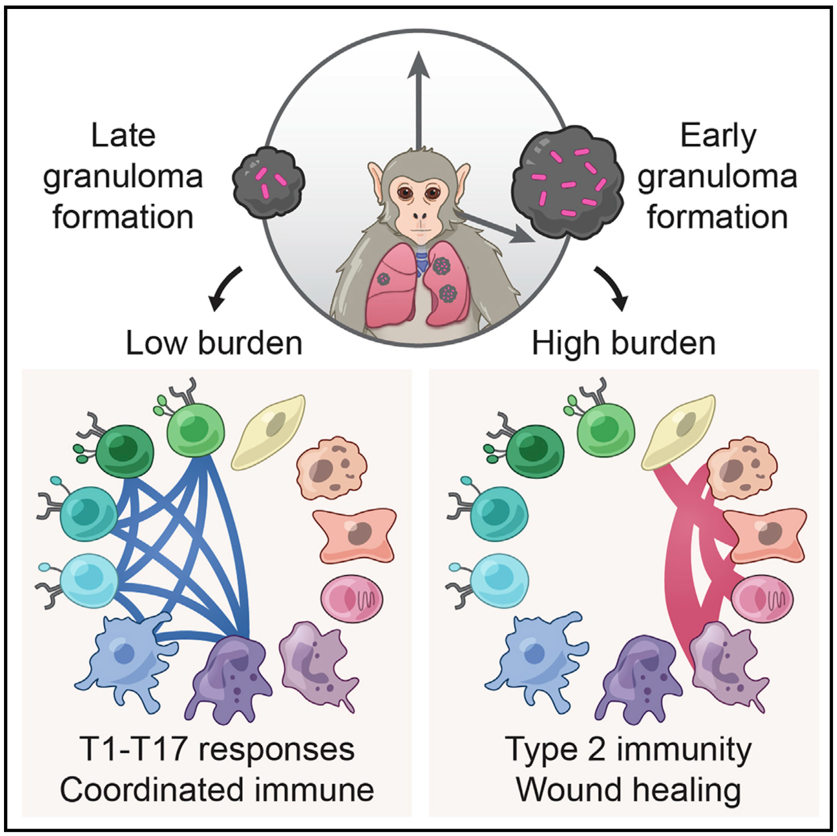
To help address this, we have established and enabled multi-group, multi-country partnerships to deploy and adapt cutting-edge genomic tools. By examining how cells dynamically alter their states, individually and collectively, during disease and/or its resolution in acute and chronic infections—e.g., tuberculosis, HIV/SHIV, hepatitis, malaria, leprosy, flu, SARS-CoV-2, and ebola—we have uncovered cellular and molecular features of pathogen control or pathology to potentiate or counteract, respectively. Illustratively, in tuberculosis, we identified a functional role for cytotoxic CD8 and hybrid type1-type17 T cells in control of infection in the lung and links between mast, plasma, and endothelial cell abundance (type-2 immune responses) and bacterial burden. We have also built methods for examining pathogens within individual host cells to define their dynamic interdependence and identify potentially restrictive host factors.

We are currently working to identify the drivers of common host responses to distinct perturbations and their targetability, as well as the impact of different interventions (e.g., vaccines).

Immune responses play a critical role in preventing tumorigenesis. Sometimes, however, they are ineffectual and can even drive/support malignancy.

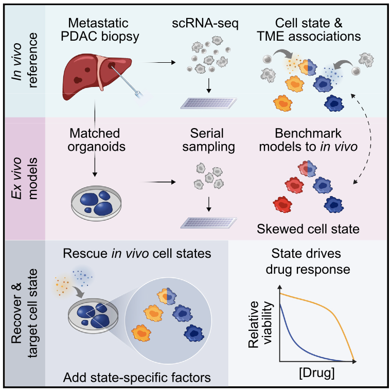
We have examined how cancer cells alter and are influenced by their tumor microenvironments (TMEs), and the impact this has on therapeutic responses. Illustratively, in Pancreatic Ductal Adenocarcinoma (PDAC), by profiling liver metastases and matched organoid models, we showed: 1. associations between TME and malignant cell state composition; 2. that autocrine and paracrine signaling can drive malignant cell state transitions, even in an isogenic background, altering the efficacy of frontline chemotherapies; and, 3. that microenvironmental manipulations can be used to control malignant state, and thereby drug responses, rationally, and to improve model fidelity for screening potential therapies. This and related work highlight the potential utility of modulating indirect target cells (T cells in the PDAC TME or basal cells in allergic inflammation) to enhance cures and preventions. 

We are now systematically expanding this work to define how additional environmental and cell-intrinsic factors influence malignant cell state plasticity in PDAC and other cancers toward enhancing treatments.


We are exposed to a constant flux of external biochemical and physical stimuli as we age. Despite variability in our overall experiences and exact constitutions, our individual tissues typically manage to maintain functionality, though each can differ in its resilience to distinct stressors.

We have characterized how differences in cellular composition and communication impact tissue fitness and have identified responses and subsequent adaptations that drive chronic dysfunction. For example, although aberrant immune activity can precipitate allergic inflammatory diseases, therapies targeting immune cells and signaling are only successful in some, suggesting chronicity may involve alternative mechanisms. Previously, we helped demonstrate that dysregulated type-2 immune signaling, driven by environmental allergens, can impact tissue health in the upper airway through generating dysfunctional basal epithelial stem cells. These stem cells can then contribute to persistence by serving as repositories for allergic inflammatory memories, altering the integrity and functional output of the nasal epithelium. Our work, with that of others, suggests generalizable principles for cellular memory, and informs where and how tissues should be targeted to support health or restore function. We have since further investigated how tissue-resident cellular subsets participate in, and are shaped by, environmental exposures at barrier tissues and the functional consequences of these experiences.

We are now working to develop a more holistic appreciation for how different intra- and extracellular factors (e.g., genetics and integrated exposure history, respectively) influence barrier tissue function.

HIV-1–specific broadly neutralizing antibodies (bnAbs) typically develop in individuals with continuous high-level viral replication and increased immune activation, conditions that cannot be reproduced during prophylactic immunization. Understanding mechanisms supporting bnAb development in the absence of high-level viremia may be important for designing bnAb-inducing immunogens. Here, we show that the breadth of neutralizing antibody responses in HIV-1 controllers was associated with a relative enrichment of circulating CXCR5+CXCR3+PD-1lo CD4+ T cells. These CXCR3+PD-1lo Tfh-like cells were preferentially induced in vitro by functionally superior dendritic cells from controller neutralizers, and able to secrete IL-21 and support B cells. In addition, these CXCR3+PD-1lo Tfh-like cells contained higher proportions of stem cell–like memory T cells, and upon antigenic stimulation differentiated into PD-1hi Tfh-like cells in a Notch-dependent manner. Together, these data suggest that CXCR5+CXCR3+PD-1lo cells represent a dendritic cell–primed precursor cell population for PD-1hi Tfh-like cells that may contribute to the generation of bnAbs in the absence of high-level viremia.

CD8+ T cell recognition of virus-infected cells is characteristically restricted by major histocompatibility complex (MHC) class I, although rare examples of MHC class II restriction have been reported in Cd4-deficient mice and a macaque SIV vaccine trial using a recombinant cytomegalovirus vector. Here, we demonstrate the presence of human leukocyte antigen (HLA) class II-restricted CD8+ T cell responses with antiviral properties in a small subset of HIV-infected individuals. In these individuals, T cell receptor β (TCRβ) analysis revealed that class II-restricted CD8+ T cells underwent clonal expansion and mediated killing of HIV-infected cells. In one case, these cells comprised 12% of circulating CD8+ T cells, and TCRα analysis revealed two distinct co-expressed TCRα chains, with only one contributing to binding of the class II HLA-peptide complex. These data indicate that class II-restricted CD8+ T cell responses can exist in a chronic human viral infection, and may contribute to immune control.

Innate lymphoid cells (ILCs) play a central role in the response to infection by secreting cytokines crucial for immune regulation, tissue homeostasis, and repair. Although dysregulation of these systems is central to pathology, the impact of HIV-1 on ILCs remains unknown. We found that human blood ILCs were severely depleted during acute viremic HIV-1 infection and that ILC numbers did not recover after resolution of peak viremia. ILC numbers were preserved by antiretroviral therapy (ART), but only if initiated during acute infection. Transcriptional profiling during the acute phase revealed upregulation of genes associated with cell death, temporally linked with a strong IFN acutephase response and evidence of gut barrier breakdown. We found no evidence of tissue redistribution in chronic disease and remaining circulating ILCs were activated but not apoptotic. These data provide a potential mechanistic link between acute HIV-1 infection, lymphoid tissue breakdown, and persistent immune dysfunction.

We introduce a microfluidic platform that enables off-chip single-cell RNA-seq after multi-generational lineage tracking under controlled culture conditions. We use this platform to generate whole-transcriptome profiles of primary, activated murine CD8+ T-cell and lymphocytic leukemia cell line lineages. Here we report that both cell types have greater intra- than inter-lineage transcriptional similarity. For CD8+ T-cells, genes with functional annotation relating to lymphocyte differentiation and function—including Granzyme B—are enriched among the genes that demonstrate greater intra-lineage expression level similarity. Analysis of gene expression covariance with matched measurements of time since division reveals cell type-specific transcriptional signatures that correspond with cell cycle progression. We believe that the ability to directly measure the effects of lineage and cell cycle-dependent transcriptional profiles of single cells will be broadly useful to fields where heterogeneous populations of cells display distinct clonal trajectories, including immunology, cancer, and developmental biology.

Extensive cellular heterogeneity exists within specific immune-cell subtypes classified as a single lineage, but its molecular underpinnings are rarely characterized at a genomic scale. Here, we use single-cell RNA-seq to investigate the molecular mechanisms governing heterogeneity and pathogenicity of Th17 cells isolated from the central nervous system (CNS) and lymph nodes (LN) at the peak of autoimmune encephalomyelitis (EAE) or differentiated in vitro under either pathogenic or non-pathogenic polarization conditions. Computational analysis relates a spectrum of cellular states in vivo to in-vitro-differentiated Th17 cells and unveils genes governing pathogenicity and disease susceptibility. Using knockout mice, we validate four new genes: Gpr65PlzpToso, and Cd5l (in a companion paper). Cellular heterogeneity thus informs Th17 function in autoimmunity and can identify targets for selective suppression of pathogenic Th17 cells while potentially sparing non-pathogenic tissue-protective ones.

Epigenetic changes are crucial for the generation of immunological memory. Failure to generate or maintain these changes will result in poor memory responses. Similarly, augmenting or stabilizing the correct epigenetic states offers a potential method of enhancing memory. Yet the transcription factors that regulate these processes are poorly defined. We find that the transcription factor Oct1 and its cofactor OCA-B are selectively required for the in vivo generation of CD4+ memory T cells. More importantly, the memory cells that are formed do not respond properly to antigen reencounter. In vitro, both proteins are required to maintain a poised state at the Il2 target locus in resting but previously stimulated CD4+ T cells. OCA-B is also required for the robust reexpression of multiple other genes including Ifng. ChIPseq identifies ∼50 differentially expressed direct Oct1 and OCA-B targets. We identify an underlying mechanism involving OCA-B recruitment of the histone lysine demethylase Jmjd1a to targets such as Il2Ifng, andZbtb32. The findings pinpoint Oct1 and OCA-B as central mediators of CD4+ T cell memory.

Encounters between immune cells and invading bacteria ultimately determine the course of infection. These interactions are usually measured in populations of cells, masking cell-to-cell variation that may be important for infection outcome. To characterize the gene expression variation that underlies distinct infection outcomes and monitor infection phenotypes, we developed an experimental system that combines single-cell RNA-seq with fluorescent markers. Probing the responses of individual macrophages to invading Salmonella, we find that variation between individual infected host cells is determined by the heterogeneous activity of bacterial factors in individual infecting bacteria. We illustrate how variable PhoPQ activity in the population of invading bacteria drives variable host type I IFN responses by modifying LPS in a subset of bacteria. This work demonstrates a causative link between host and bacterial variability, with cell-to-cell variation between different bacteria being sufficient to drive radically different host immune responses. This co-variation has implications for host-pathogen dynamics in vivo.

The mammalian immune system is tasked with protecting the host against a broad range of threats. Understanding how immune populations leverage cellular diversity to achieve this breadth and flexibility, particularly during dynamic processes such as differentiation and antigenic response, is a core challenge that is well suited for single cell analysis. Recent years have witnessed transformative and intersecting advances in nanofabrication and genomics that enable deep profiling of individual cells, affording exciting opportunities to study heterogeneity in the immune response at an unprecedented scope. In light of these advances, here we review recent work exploring how immune populations generate and leverage cellular heterogeneity at multiple molecular and phenotypic levels. Additionally, we highlight opportunities for single cell technologies to shed light on the causes and consequences of heterogeneity in the immune system.

Recent molecular studies have shown that, even when derived from a seemingly homogenous population, individual cells can exhibit substantial differences in gene expression, protein levels and phenotypic output1–5, with important functional consequences4,5. Existing studies of cellular heterogeneity, however, have typically measured only a few pre-selected RNAs1,2 or proteins5,6 simultaneously, because genomic profiling methods3 could not be applied to single cells until very recently7–10. Here we use single-cell RNA sequencing to investigate heterogeneity in the response of mouse bone-marrow-derived dendritic cells (BMDCs) to lipopolysaccharide. We find extensive, and previously unobserved, bimodal variation in messenger RNA abundance and splicing patterns, which we validate by RNA-fluorescence in situ hybridization for select transcripts. In particular, hundreds of key immune genes are bimodally expressed across cells, surprisingly even for genes that are very highly expressed at the population average. Moreover, splicing patterns demonstrate previously unobserved levels of heterogeneity between cells. Some of the observed bimodality can be attributed to closely related, yet distinct, known maturity states of BMDCs; other portions reflect differences in the usage of key regulatory circuits. For example, we identify a module of 137 highly variable, yet co-regulated, antiviral response genes. Using cells from knockout mice, we show that variability in this module may be propagated through an interferon feedback circuit, involving the transcriptional regulators Stat2 and Irf7. Our study demonstrates the power and promise of single-cell genomics in uncovering functional diversity between cells and in deciphering cell states and circuits.

Individual genetic variation affects gene responsiveness to stimuli, often by influencing complex molecular circuits. Here we combine genomic and intermediate-scale transcriptional profiling with computational methods to identify variants that affect the responsiveness of genes to stimuli (responsiveness quantitative trait loci or reQTLs) and to position these variants in molecular circuit diagrams. We apply this approach to study variation in transcriptional responsiveness to pathogen components in dendritic cells from recombinant inbred mouse strains. We identify reQTLs that correlate with particular stimuli and position them in known pathways. For example, in response to a virus-like stimulus, a trans-acting variant responds as an activator of the antiviral response; using RNA interference, we identify Rgs16 as the likely causal gene. Our approach charts an experimental and analytic path to decipher the mechanisms underlying genetic variation in circuits that control responses to stimuli.

Despite their importance, the molecular circuits that control the differentiation of naive T cells remain largely unknown. Recent studies that reconstructed regulatory networks in mammalian cells have focused on short-term responses and relied on perturbation-based approaches that cannot be readily applied to primary T cells. Here we combine transcriptional profiling at high temporal resolution, novel computational algorithms, and innovative nanowirebased perturbation tools to systematically derive and experimentally validate a model of the dynamic regulatory network that controls the differentiation of mouse TH17 cells, a proinflammatory T-cell subset that has been implicated in the pathogenesis of multiple autoimmune diseases. The TH17 transcriptional network consists of two self-reinforcing, but mutually antagonistic, modules, with 12 novel regulators, the coupled action of which may be essential for maintaining the balance between TH17 and other CD4+ T-cell subsets. Our study identifies and validates 39 regulatory factors, embeds them within a comprehensive temporal network and reveals its organizational principles; it also highlights novel drug targets for controlling TH17 cell differentiation.

Developing a detailed understanding of enzyme function in the context of an intracellular signal transduction pathway requires minimally invasive methods for probing enzyme activity in situ. Here, we describe a new method for monitoring enzyme activity in living cells by sandwiching live cells between two vertical silicon nanowire (NW) arrays. Specifically, we use the first NW array to immobilize the cells and then present enzymatic substrates intracellularly via the second NW array by utilizing the NWs’ ability to penetrate cellular membranes without affecting cells’ viability or function. This strategy, when coupled with fluorescence microscopy and mass spectrometry, enables intracellular examination of protease, phosphatase, and protein kinase activities, demonstrating the assay’s potential in uncovering the physiological roles of various enzymes.

Deciphering the signaling networks that underlie normal and disease processes remains a major challenge. Here, we report the discovery of signaling components involved in the Toll-like receptor (TLR) response of immune dendritic cells (DCs), including a previously unkown pathway shared across mammalian antiviral responses. By combining transcriptional profiling, genetic and small-molecule perturbations, and phosphoproteomics, we uncover 35 signaling regulators, including 16 known regulators, involved in TLR signaling. In particular, we find that Polo-like kinases (Plk) 2 and 4 are essential components of antiviral pathways in vitro and in vivo and activate a signaling branch involving a dozen proteins, among which is Tnfaip2, a gene associated with autoimmune diseases but whose role was unknown. Our study illustrates the power of combining systematic measurements and perturbations to elucidate complex signaling circuits and discover potential therapeutic targets.