Single-Cell Analyses of Colon and Blood Reveal Distinct Immune Cell Signatures of Ulcerative Colitis and Crohn’s Disease
 Biology
 Genomics
 Immunology
 Alex K. Shalek
 José Ordovas-Montañes
 Marko Vukovic
Gastroenterology , Volume 159
September, 2020
Abstract
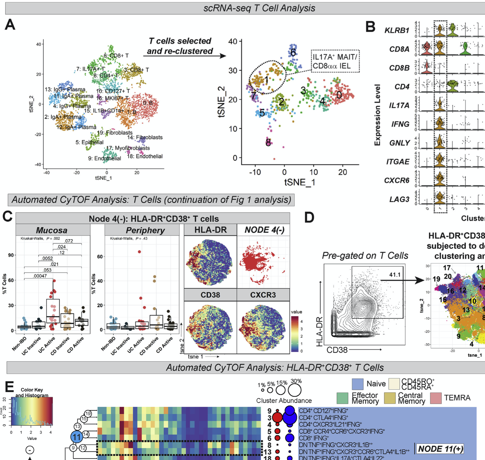
Studies are needed to determine the mechanisms of mucosal dysregulation in patients with inflammatory bowel diseases (IBDs) and differences in inflammatory responses of patients with ulcerative colitis (UC) vs Crohn’s disease (CD). We used mass cytometry (CyTOF) to characterize and compare immune cell populations in the mucosa and blood from patients with IBD and without IBD (controls) at single-cell resolution. We performed CyTOF analysis of colonic mucosa samples (n = 87) and peripheral blood mononuclear cells (n = 85) from patients with active or inactive UC or CD and controls. We also performed single-cell RNA sequencing, flow cytometry, and RNA in situ hybridization analyses to validate key findings. We used random forest modeling to identify differences in signatures across subject groups. Compared with controls, colonic mucosa samples from patients with IBD had increased abundances of HLA-DR+CD38+ T cells, including T-regulatory cells that produce inflammatory cytokines; CXCR3+ plasmablasts; and IL1B+ macrophages and monocytes. Colonic mucosa samples from patients with UC were characterized by expansion of IL17A+ CD161+ effector memory T cells and IL17A+ T-regulatory cells; expansion of HLA-DR+CD56+ granulocytes; and reductions in type 3 innate lymphoid cells. Mucosal samples from patients with active CD were characterized by IL1B+HLA-DR+CD38+ T cells, IL1B+TNF+IFNG+ naïve B cells, IL1B+ dendritic cells (DCs), and IL1B+ plasmacytoid DCs. Peripheral blood mononuclear cells from patients with active CD differed from those of active UC in that the peripheral blood mononuclear cells from patients with CD had increased IL1B+ T-regulatory cells, IL1B+ DCs and IL1B+ plasmacytoid DCs, IL1B+ monocytes, and fewer group 1 innate lymphoid cells. Random forest modeling differentiated active UC from active CD in colonic mucosa and blood samples; top discriminating features included many of the cellular populations identified above. We used single-cell technologies to identify immune cell populations specific to mucosa and blood samples from patients with active or inactive CD and UC and controls. This information might be used to develop therapies that target specific cell populations in patients with different types of IBD.