Computational cohort materials from March 2023 Ghana training

Imidazoquinolines (IMDs), such as resiquimod (R848), are of great interest as potential cancer immunotherapies because of their ability to activate Toll-like receptor 7 (TLR7) and/or TLR8 on innate immune cells. Nevertheless, intravenous administration of IMDs causes severe immune-related toxicities, and attempts to improve their tissue-selective exposure while minimizing acute systemic inflammation have proven difficult. Here, using a library of R848 “bottlebrush prodrugs” (BPDs) that differ only by their R848 release kinetics, we explore how the timing of R848 exposure affects immune stimulation in vitro and in vivo. These studies led to the discovery of R848-BPDs that exhibit optimal activation kinetics to achieve potent stimulation of myeloid cells in tumors and substantial reductions in tumor growth following systemic administration in mouse syngeneic tumor models without any observable systemic toxicity. These results suggest that release kinetics can be tuned at the molecular level to provide safe yet effective systemically administered immunostimulant prodrugs for next-generation cancer immunotherapies.

Loss of the PTEN tumour suppressor is one of the most common oncogenic drivers across all cancer types. PTEN is the major negative regulator of PI3K signalling. The PI3Kβ isoform has been shown to play an important role in PTEN-deficient tumours, but the mechanisms underlying the importance of PI3Kβ activity remain elusive. Here, using a syngeneic genetically engineered mouse model of invasive breast cancer driven by ablation of both Ptenand Trp53 (which encodes p53), we show that genetic inactivation of PI3Kβ led to a robust anti-tumour immune response that abrogated tumour growth in syngeneic immunocompetent mice, but not in immunodeficient mice. Mechanistically, PI3Kβ inactivation in the PTEN-null setting led to reduced STAT3 signalling and increased the expression of immune stimulatory molecules, thereby promoting anti-tumour immune responses. Pharmacological PI3Kβ inhibition also elicited anti-tumour immunity and synergized with immunotherapy to inhibit tumour growth. Mice with complete responses to the combined treatment displayed immune memory and rejected tumours upon re-challenge. Our findings demonstrate a molecular mechanism linking PTEN loss and STAT3 activation in cancer and suggest that PI3Kβ controls immune escape in PTEN-null tumours, providing a rationale for combining PI3Kβ inhibitors with immunotherapy for the treatment of PTEN-deficient breast cancer.

Chronic liver disease and hepatocellular carcinoma (HCC) are life-threatening diseases with limited treatment options. The lack of clinically relevant/tractable experimental models hampers therapeutic discovery. Here, we develop a simple and robust human liver cell-based system modeling a clinical prognostic liver signature (PLS) predicting long-term liver disease progression toward HCC. Using the PLS as a readout, followed by validation in nonalcoholic steatohepatitis/fibrosis/HCC animal models and patient-derived liver spheroids, we identify nizatidine, a histamine receptor H2 (HRH2) blocker, for treatment of advanced liver disease and HCC chemoprevention. Moreover, perturbation studies combined with single cell RNA-Seq analyses of patient liver tissues uncover hepatocytes and HRH2+, CLEC5Ahigh, MARCOlowliver macrophages as potential nizatidine targets. The PLS model combined with single cell RNA-Seq of patient tissues enables discovery of urgently needed targets and therapeutics for treatment of advanced liver disease and cancer prevention.

High-throughput phenotypic screens leveraging biochemical perturbations, high-content readouts, and complex multicellular models could advance therapeutic discovery yet remain constrained by limitations of scale. To address this, we establish a method for compressing screens by pooling perturbations followed by computational deconvolution. Conducting controlled benchmarks with a highly bioactive small molecule library and a high-content imaging readout, we demonstrate increased efficiency for compressed experimental designs compared to conventional approaches. To prove generalizability, we apply compressed screening to examine transcriptional responses of patient-derived pancreatic cancer organoids to a library of tumor-microenvironment (TME)-nominated recombinant protein ligands. Using single-cell RNA-seq as a readout, we uncover reproducible phenotypic shifts induced by ligands that correlate with clinical features in larger datasets and are distinct from reference signatures available in public databases. In sum, our approach enables phenotypic screens that interrogate complex multicellular models with rich phenotypic readouts to advance translatable drug discovery as well as basic biology.

Pancreatic ductal adenocarcinoma (PDAC) has been classified into classical and basal-like transcriptional subtypes by bulk RNA measurements. However, recent work has uncovered greater complexity to transcriptional subtypes than was initially appreciated using bulk RNA expression profiling. To provide a deeper understanding of PDAC subtypes, we developed a multiplex immunofluorescence (mIF) pipeline that quantifies protein expression of six PDAC subtype markers (CLDN18.2, TFF1, GATA6, KRT17, KRT5, and S100A2) and permits spatially resolved, single-cell interrogation of pancreatic tumors from resection specimens and core needle biopsies. Both primary and metastatic tumors displayed striking intratumoral subtype heterogeneity that was associated with patient outcomes, existed at the scale of individual glands, and was significantly reduced in patient-derived organoid cultures. Tumor cells co-expressing classical and basal markers were present in > 90% of tumors, existed on a basal-classical polarization continuum, and were enriched in tumors containing a greater admixture of basal and classical cell populations. Cell-cell neighbor analyses within tumor glands further suggested that co-expressor cells may represent an intermediate state between expression subtype poles. The extensive intratumoral heterogeneity identified through this clinically applicable mIF pipeline may inform prognosis and treatment selection for patients with PDAC.

High-risk forms of B-acute lymphoblastic leukemia (B-ALL) remain a therapeutic challenge. Leukemia-initiating cells (LICs) self-renew and spark relapse and therefore have been the subject of intensive investigation; however, the properties of LICs in high-risk B-ALL are not well understood. Here, we use single-cell transcriptomics and quantitative xenotransplantation to understand LICs in MLL-rearranged (MLL-r) B-ALL. Compared with reported LIC frequencies in acute myeloid leukemia (AML), engraftable LICs in MLL-r B-ALL are abundant. Although we find that multipotent, self-renewing LICs are enriched among phenotypically undifferentiated B-ALL cells, LICs with the capacity to replenish the leukemic cellular diversity can emerge from more mature fractions. While inhibiting oxidative phosphorylation blunts blast proliferation, this intervention promotes LIC emergence. Conversely, inhibiting hypoxia and glycolysis impairs MLL-r B-ALL LICs, providing a therapeutic benefit in xenotransplantation systems. These findings provide insight into the aggressive nature of MLL-r B-ALL and provide a rationale for therapeutic targeting of hypoxia and glycolysis.

The immune system represents a major barrier to cancer progression, driving the evolution of immunoregulatory interactions between malignant cells and T-cells in the tumor environment. Blastic plasmacytoid dendritic cell neoplasm (BPDCN), a rare acute leukemia with plasmacytoid dendritic cell (pDC) differentiation, provides a unique opportunity to study these interactions. pDCs are key producers of interferon alpha (IFNA) that play an important role in T-cell activation at the interface between the innate and adaptive immune system. To assess how uncontrolled proliferation of malignant BPDCN cells affects the tumor environment, we catalog immune cell heterogeneity in the bone marrow (BM) of five healthy controls and five BPDCN patients by analyzing 52,803 single-cell transcriptomes, including 18,779 T-cells. We test computational techniques for robust cell type classification and find that T-cells in BPDCN patients consistently upregulate interferon alpha (IFNA) response and downregulate tumor necrosis factor alpha (TNFA) pathways. Integrating transcriptional data with T-cell receptor sequencing via shared barcodes reveals significant T-cell exhaustion in BPDCN that is positively correlated with T-cell clonotype expansion. By highlighting new mechanisms of T-cell exhaustion and immune evasion in BPDCN, our results demonstrate the value of single-cell multiomics to understand immune cell interactions in the tumor environment.

Oncogenes act in a cell-intrinsic way to promote tumorigenesis. Whether oncogenes also have a cell-extrinsic effect on suppressing the immune response to cancer is less well understood. We use an in vivo expression screen of known cancer-associated somatic mutations in mouse syngeneic tumor models treated with checkpoint blockade to identify oncogenes that promote immune evasion. We then validated candidates from this screen in vivo and analyzed the tumor immune microenvironment of tumors expressing mutant protein to identify mechanisms of immune evasion. We found that expression of a catalytically active mutation in phospho-inositol 3 kinase (PI3K), PIK3CA c.3140A>G (H1047R) confers a selective growth advantage to tumors treated with immunotherapy that is reversed by pharmacological PI3K inhibition. PIK3CA H1047R-expression in tumors decreased the number of CD8+ T cells but increased the number of inhibitory myeloid cells following immunotherapy. Inhibition of myeloid infiltration by pharmacological or genetic modulation of Ccl2 in PIK3CA H1047R tumors restored sensitivity to programmed cell death protein 1 (PD-1) checkpoint blockade. PI3K activation enables tumor immune evasion by promoting an inhibitory myeloid microenvironment. Activating mutations in PI3K may be useful as a biomarker of poor response to immunotherapy. Our data suggest that some oncogenes promote tumorigenesis by enabling tumor cells to avoid clearance by the immune system. Identification of those mechanisms can advance rational combination strategies to increase the efficacy of immunotherapy.

Immunosurveillance of cancer requires the presentation of peptide antigens on major histocompatibility complex class I (MHC-I) molecules1–5. Current approaches to profiling of MHC-I-associated peptides, collectively known as the immunopeptidome, are limited to in vitro investigation or bulk tumour lysates, which limits our understanding of cancer-specific patterns of antigen presentation in vivo6. To overcome these limitations, we engineered an inducible affinity tag into the mouse MHC-I gene (H2-K1) and targeted this allele to the KrasLSL-G12D/+Trp53fl/fl mouse model (KP/KbStrep)7. This approach enabled us to precisely isolate MHC-I peptides from autochthonous pancreatic ductal adenocarcinoma and from lung adenocarcinoma (LUAD) in vivo. In addition, we profiled the LUAD immunopeptidome from the alveolar type 2 cell of origin up to late-stage disease. Differential peptide presentation in LUAD was not predictable by mRNA expression or translation efficiency and is probably driven by post-translational mechanisms. Vaccination with peptides presented by LUAD in vivo induced CD8+ T cell responses in naive mice and tumour-bearing mice. Many peptides specific to LUAD, including immunogenic peptides, exhibited minimal expression of the cognate mRNA, which prompts the reconsideration of antigen prediction pipelines that triage peptides according to transcript abundance8. Beyond cancer, the KbStrep allele is compatible with other Cre-driver lines to explore antigen presentation in vivo in the pursuit of understanding basic immunology, infectious disease and autoimmunity.

Prostate cancer is the second most common malignancy in men worldwide and consists of a mixture of tumor and non-tumor cell types. To characterize the prostate cancer tumor microenvironment, we perform single-cell RNA-sequencing on prostate biopsies, prostatectomy specimens, and patient-derived organoids from localized prostate cancer patients. We uncover heterogeneous cellular states in prostate epithelial cells marked by high androgen signaling states that are enriched in prostate cancer and identify a population of tumor-associated club cells that may be associated with prostate carcinogenesis. ERG-negative tumor cells, compared to ERG-positive cells, demonstrate shared heterogeneity with surrounding luminal epithelial cells and appear to give rise to common tumor microenvironment responses. Finally, we show that prostate epithelial organoids harbor tumor-associated epithelial cell states and are enriched with distinct cell types and states from their parent tissues. Our results provide diagnostically relevant insights and advance our understanding of the cellular states associated with prostate carcinogenesis.

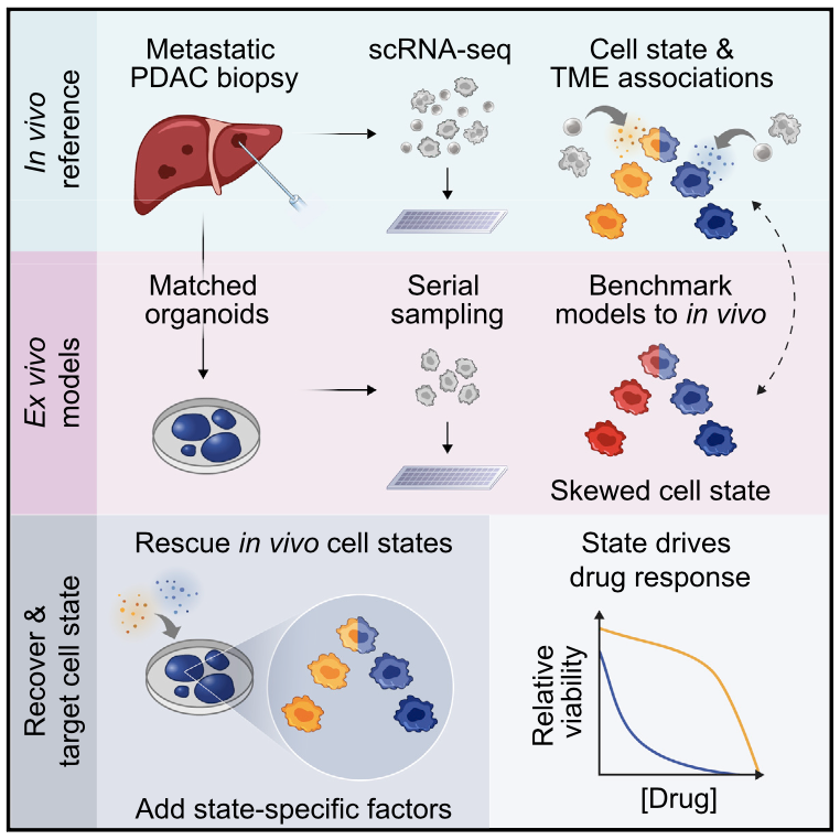
Prognostically relevant RNA expression states exist in pancreatic ductal adenocarcinoma (PDAC), but our understanding of their drivers, stability, and relationship to therapeutic response is limited. To examine these attributes systematically, we profiled metastatic biopsies and matched organoid models at single-cell resolution. In vivo, we identify a new intermediate PDAC transcriptional cell state and uncover distinct site- and state-specific tumor microenvironments (TMEs). Benchmarking models against this reference map, we reveal strong culture-specific biases in cancer cell transcriptional state representation driven by altered TME signals. We restore expression state heterogeneity by adding back in vivo-relevant factors and show plasticity in culture models. Further, we prove that non-genetic modulation of cell state can strongly influence drug responses, uncovering state-specific vulnerabilities. This work provides a broadly applicable framework for aligning cell states across in vivo and ex vivo settings, identifying drivers of transcriptional plasticity and manipulating cell state to target associated vulnerabilities.

Existing preclinical methods for acquiring dissemination kinetics of rare circulating tumor cells (CTCs) en route to forming metastases have not been capable of providing a direct measure of CTC intravasation rate and subsequent half-life in the circulation. Here, we demonstrate an approach for measuring endogenous CTC kinetics by continuously exchanging CTC-containing blood over several hours between un-anesthetized, tumor-bearing mice and healthy, tumor-free counterparts. By tracking CTC transfer rates, we extrapolated half-life times in the circulation of between 40 and 260 s and intravasation rates between 60 and 107,000 CTCs/hour in mouse models of small-cell lung cancer (SCLC), pancreatic ductal adenocarcinoma (PDAC), and non-small cell lung cancer (NSCLC). Additionally, direct transfer of only 1−2% of daily-shed CTCs using our blood-exchange technique from late-stage, SCLC-bearing mice generated macrometastases in healthy recipient mice. We envision that our technique will help further elucidate the role of CTCs and the rate-limiting steps in metastasis.

Leptomeningeal disease (LMD) is a devastating complication of solid tumor malignancies, with dire prognosis and no effective systemic treatment options. Over the past decade, the incidence of LMD has steadily increased due to therapeutics that have extended the survival of cancer patients, highlighting the need for new interventions. To examine the efficacy of immune checkpoint inhibitors (ICI) in patients with LMD, we completed two phase II clinical trials. Here, we investigate the cellular and molecular features underpinning observed patient trajectories in these trials by applying single-cell RNA and cell-free DNA profiling to longitudinal cerebrospinal fluid (CSF) draws from enrolled patients. We recover immune and malignant cell types in the CSF, characterize cell behavior changes following ICI, and identify genomic features associated with relevant clinical phenomena. Overall, our study describes the liquid LMD tumor microenvironment prior to and following ICI treatment and demonstrates clinical utility of cell-free and single-cell genomic measurements for LMD research.

Immune checkpoint blockers (ICBs) have failed in all phase III glioblastoma (GBM) trials. Here, we show that regulatory T (Treg) cells play a key role in GBM resistance to ICBs in experimental gliomas. Targeting glucocorticoid-induced TNFR-related receptor (GITR) in Treg cells using an agonistic antibody (αGITR) promotes CD4 Treg cell differentiation into CD4 effector T cells, alleviates Treg cell-mediated suppression of anti-tumor immune response, and induces potent anti-tumor effector cells in GBM. The reprogrammed GBM-infiltrating Treg cells express genes associated with a Th1 response signature, produce IFNγ, and acquire cytotoxic activity against GBM tumor cells while losing their suppressive function. αGITR and αPD1 antibodies increase survival benefit in three experimental GBM models, with a fraction of cohorts exhibiting complete tumor eradication and immune memory upon tumor re-challenge. Moreover, αGITR and αPD1 synergize with the standard of care treatment for newly-diagnosed GBM, enhancing the cure rates in these GBM models.

Central to anti-tumor immunity are dendritic cells (DCs), which stimulate long-lived protective T cell responses. Recent studies have demonstrated that DCs can achieve a state of hyperactivation, which is associated with inflammasome activities within living cells. Herein, we report that hyperactive DCs have an enhanced ability to migrate to draining lymph nodes and stimulate potent cytotoxic T lymphocyte (CTL) responses. This enhanced migratory activity is dependent on the chemokine receptor CCR7 and is associated with a unique transcriptional program that is not observed in conventionally activated or pyroptotic DCs. We show that hyperactivating stimuli are uniquely capable of inducing durable CTL-mediated anti-tumor immunity against tumors that are sensitive or resistant to PD-1 inhibition. These protective responses are intrinsic to the cDC1 subset of DCs, depend on the inflammasome-dependent cytokine IL-1β, and enable tumor lysates to serve as immunogens. If these activities are verified in humans, hyperactive DCs may impact immunotherapy.

Prostate cancer is the second most common malignancy in men worldwide and consists of a mixture of tumor and non-tumor cell types. To characterize the prostate cancer tumor microenvironment, we performed single-cell RNA-sequencing on prostate biopsies, prostatectomy specimens, and patient-derived organoids from localized prostate cancer patients. We identify a population of tumor-associated club cells that may act as progenitor cells and uncover heterogeneous cellular states in prostate epithelial cells marked by high androgen signaling states that are enriched in prostate cancer. ERG- tumor cells, compared to ERG+ cells, demonstrate shared heterogeneity with surrounding luminal epithelial cells and appear to give rise to common tumor microenvironment responses. Finally, we show that prostate epithelial organoids recapitulate tumor-associated epithelial cell states and are enriched with distinct cell types and states from their parent tissues. Our results provide diagnostically relevant insights and advance our understanding of the cellular states associated with prostate carcinogenesis.

Bulk transcriptomic studies have defined classical and basal-like gene expression subtypes in pancreatic ductal adenocarcinoma (PDAC) that correlate with survival and response to chemotherapy; however, the underlying mechanisms that govern these subtypes and their heterogeneity remain elusive. Here, we performed single-cell RNA-sequencing of 23 metastatic PDAC needle biopsies and matched organoid models to understand how tumor cell-intrinsic features and extrinsic factors in the tumor microenvironment (TME) shape PDAC cancer cell phenotypes. We identify a novel cancer cell state that co-expresses basal-like and classical signatures, demonstrates upregulation of developmental and KRAS-driven gene expression programs, and represents a transitional intermediate between the basal-like and classical poles. Further, we observe structure to the metastatic TME supporting a model whereby reciprocal intercellular signaling shapes the local microenvironment and influences cancer cell transcriptional subtypes. In organoid culture, we find that transcriptional phenotypes are plastic and strongly skew toward the classical expression state, irrespective of genotype. Moreover, we show that patient-relevant transcriptional heterogeneity can be rescued by supplementing organoid media with factors found in the TME in a subtype-specific manner. Collectively, our study demonstrates that distinct microenvironmental signals are critical regulators of clinically relevant PDAC transcriptional states and their plasticity, identifies the necessity for considering the TME in cancer modeling efforts, and provides a generalizable approach for delineating the cell-intrinsic versus -extrinsic factors that govern tumor cell phenotypes.

Crucial transitions in cancer—including tumor initiation, local expansion, metastasis, and therapeutic resistance—involve complex interactions between cells within the dynamic tumor ecosystem. Transformative single-cell genomics technologies and spatial multiplex in situ methods now provide an opportunity to interrogate this complexity at unprecedented resolution. The Human Tumor Atlas Network (HTAN), part of the National Cancer Institute (NCI) Cancer Moonshot Initiative, will establish a clinical, experimental, computational, and organizational framework to generate informative and accessible three-dimensional atlases of cancer transitions for a diverse set of tumor types. This effort complements both ongoing efforts to map healthy organs and previous large-scale cancer genomics approaches focused on bulk sequencing at a single point in time. Generating single-cell, multiparametric, longitudinal atlases and integrating them with clinical outcomes should help identify novel predictive biomarkers and features as well as therapeutically relevant cell types, cell states, and cellular interactions across transitions. The resulting tumor atlases should have a profound impact on our understanding of cancer biology and have the potential to improve cancer detection, prevention, and therapeutic discovery for better precision-medicine treatments of cancer patients and those at risk for cancer.

Whether cultured in vitro or part of a complex tissue in vivo, a cell’s phenotype and function are significantly influenced by dynamic interactions with its microenvironment. To explicitly examine how a cell’s spatiotemporal activity impacts its behavior, we developed and validated a strategy termed SPACECAT – Spatially PhotoActivatable Color Encoded Cell Address Tags – to annotate, track, and isolate specific cells in a non-destructive, viability-preserving manner. In SPACECAT, a biological sample is immersed in a photocaged fluorescent molecule, and cells within a location of interest are labeled for further study by uncaging that molecule with user-patterned near-UV light. SPACECAT offers high spatial precision and temporal stability across diverse cell and tissue types, and is compatible with common downstream assays, including flow cytometry and single-cell RNA-Seq. Illustratively, we leveraged this approach in patient-derived intestinal organoids, a spatially complex system less amenable to genetic manipulations, to select for crypt-like regions enriched in stem-like and actively mitotic cells. Moreover, we demonstrate its applicability and utility on ex vivo tissue sections from four healthy organs and an autochthonous lung tumor model, uncovering spatially-biased gene expression patterns among immune cell subsets and identifying rare myeloid phenotypes enriched around tumor/healthy border regions. In sum, our method provides a minimally invasive and broadly applicable approach to link cellular spatiotemporal features and/or behavioral phenotypes with diverse downstream assays, enabling fundamental insights into the connections between tissue microenvironments and biological (dys)function.

Pilocytic astrocytoma (PA), the most common childhood brain tumor, is a low-grade glioma with a single driver BRAF rearrangement. Here, we perform scRNAseq in six PAs using methods that enabled detection of the rearrangement. When compared to higher-grade gliomas, a strikingly higher proportion of the PA cancer cells exhibit a differentiated, astrocyte-like phenotype. A smaller proportion of cells exhibit a progenitor-like phenotype with evidence of proliferation. These express a mitogen-activated protein kinase (MAPK) programme that was absent from higher-grade gliomas. Immune cells, especially microglia, comprise 40% of all cells in the PAs and account for differences in bulk expression profiles between tumor locations and subtypes. These data indicate that MAPK signaling is restricted to relatively undifferentiated cancer cells in PA, with implications for investigational therapies directed at this pathway.

The extraordinary activity of high-dose cyclophosphamide against some high-grade lymphomas was described nearly 60 years ago. Here we address mechanisms that mediate cyclophosphamide activity in bona fide human double-hit lymphoma. We show that antibody resistance within the bone marrow (BM) is not present upon early engraftment but develops during lymphoma progression. This resistance required a high tumor: macrophage ratio, was recapitulated in spleen by partial macrophage depletion and was overcome by multiple, highdose alkylating agents. Cyclophosphamide induced ER-stress in BM-resident lymphoma cells in vivo that resulted in ATF4-mediated paracrine secretion of VEGF-A, massive macrophage infiltration and clearance of alemtuzumab-opsonized cells. BM macrophages isolated after cyclophosphamide treatment had increased phagocytic capacity that was reversed by VEGF-A blockade or SYK inhibition. Single-cell RNA sequencing of these macrophages identified a “super-phagocytic” subset that expressed CD36/FcgRIV. Together, these findings define a novel mechanism through which high-dose alkylating agents promote macrophage-dependent lymphoma clearance.

Circulating tumor cells (CTCs) play a fundamental role in cancer progression. However, in mice, limited blood volume and the rarity of CTCs in the bloodstream preclude longitudinal, in-depth studies of these cells using existing liquid biopsy techniques. Here, we present an optofluidic system that continuously collects fluorescently labeled CTCs from a genetically engineered mouse model (GEMM) for several hours per day over multiple days or weeks. The system is based on a microfluidic cell sorting chip connected serially to an unanesthetized mouse via an implanted arteriovenous shunt. Pneumatically controlled microfluidic valves capture CTCs as they flow through the device, and CTC-depleted blood is returned back to the mouse via the shunt. To demonstrate the utility of our system, we profile CTCs isolated longitudinally from animals over 4 days of treatment with the BET inhibitor JQ1 using single-cell RNA sequencing (scRNA-Seq) and show that our approach eliminates potential biases driven by intermouse heterogeneity that can occur when CTCs are collected across different mice. The CTC isolation and sorting technology presented here provides a research tool to help reveal details of how CTCs evolve over time, allowing studies to credential changes in CTCs as biomarkers of drug response and facilitating future studies to understand the role of CTCs in metastasis.

Mass and growth rate are highly integrative measures of cell physiology not discernable via genomic measurements. Here, we introduce a microfluidic platform enabling direct measurement of single-cell mass and growth rate upstream of highly multiplexed single-cell profiling such as single-cell RNA sequencing. We resolve transcriptional signatures associated with single-cell mass and growth rate in L1210 and FL5.12 cell lines and activated CD8+ T cells. Further, we demonstrate a framework using these linked measurements to characterize biophysical heterogeneity in a patient-derived glioblastoma cell line with and without drug treatment. Our results highlight the value of coupled phenotypic metrics in guiding single-cell genomics.

We develop single-cell transcriptomic approaches to comprehensively profile human tissues and model systems. Previously, we focused on establishingvalidating, scaling, and simplifying single-cell RNA-seq, often through the development of microdevices, to enable genome-wide identification of the cell types/states contained within complex biological samples. More recently, we helped both enhance the detection of phenotype-defining transcripts using these methods and simplify their on-site processing for clinical applications. In parallel, we have also worked to democratize these techniques, providing open access to resources and protocols, training thousands locally and abroad, and establishing infrastructure and on-site collaborations spanning across 6 continents and 26+ countries.

As many factors define cellular phenotype and influence disease beyond mRNA, we develop complementary methods for co-assaying other cellular attributes to enrich our understanding of the drivers of cellular behaviors. Examples including the abundance of additional ‘-omes’, the sequence and amount of important transcripts, cellular history, biophysical properties, spatial position, and functional output. Recently, we have worked to: 1. detect pathogens in cells and potentially actionable associated host factors; 2. query for specific mutations to identify cancer cells; and, 3. extract T cell receptor sequences to examine clonality. We have also formulated computational methods to derive deeper insights from these data (e.g., to examine viral dynamic in infected cells, reproducible features hidden by inter-individual variability, multicellular immune dynamics, intercellular communication, or alteration in cellular ecosystems associated with pathology).

We explore how the extracellular milieu influences cellular decision-making. Here, we have employed controlled culture conditions with cells and organoids, chemical and genetic perturbations, and constant microfluidic perfusion. We also have leveraged natural microenvironmental variation within and across tissues via microdissection and by using photoactivatable probes that retain spatial information through dissociation. In each instance, we aim to understand the degree to which extracellular environments modulate, and can be used to rationally control, the responses of individual cells or the overall distribution thereof, with an eye toward engineering tissue responses.

We examine the impact of intercellular interactions on cellular function. We have used coculture, imaging and perturbation strategies, as well as matched computational methods, to reinforce findings from dissociated samples, validate inferred cell-cell communication in vivo (e.g., between sensory neurons and lymph node resident cells), and manipulate multicellular systems (e.g., organoids). We are currently working on building arrayed, synthetically-designed cellular ensembles to examine how ‘tissue’ structure impacts functional response. Our overall goal is to understand cellular co-dependencies that influence niche- and tissue-level response dynamics.

We broadly study how intra- and extracellular circuits collectively drive healthy and diseased tissue states. By leveraging the massive genomic datasets we and others have generated from complex tissues (like melanoma tumors, inflamed gut, and nasal polyps), we have begun to identify common and unique cell types/states and circuits associated with pathology that may be important for regulating biological function and stability. Our current findings suggest multiple overlaps among distinct diseases, pointing to the possibility of a finite set of evolved response strategies and thus common interventions based on adjusting specific cell states, cell frequencies, and/or cell-cell communication pathways.

Homeostatic programs balance immune protection and self-tolerance. Such mechanisms likely impact autoimmunity and tumor formation, respectively. How homeostasis is maintained and impacts tumor surveillance is unknown. Here, we find that different immune mononuclear phagocytes share a conserved steady-state program during differentiation and entry into healthy tissue. IFNγ is necessary and sufficient to induce this program, revealing a key instructive role. Remarkably, homeostatic and IFNγ-dependent programs enrich across primary human tumors, including melanoma, and stratify survival. Single-cell RNA sequencing (RNA-seq) reveals enrichment of homeostatic modules in monocytes and DCs from human metastatic melanoma. Suppressor-of-cytokine-2 (SOCS2) protein, a conserved program transcript, is expressed by mononuclear phagocytes infiltrating primary melanoma and is induced by IFNγ. SOCS2 limits adaptive anti-tumoral immunity and DC-based priming of T cells in vivo, indicating a critical regulatory role. These findings link immune homeostasis to key determinants of anti-tumoral immunity and escape, revealing co-opting of tissue-specific immune development in the tumor microenvironment.

Seq-Well is a portable, low-cost platform for single-cell RNA sequencing designed to be compatible with low-input, clinical biopsies. We have recently published a manuscript detailing the development and validation of the Seq-Well plaftorm. Here, we provide an in-depth protocol and videos describing how to perform Seq-Well experiments.

We hope that you will use Seq-Well and apply it in your work.

Seq-Well: portable, low-cost RNA sequencing of single cells at high throughput
  • Gierahn et al.,
  • Nature Methods,
  • 2017
  • R&D
  • Chemistry
  • Technology
  • Travel
  • Genomics
  • Marc Wadsworth II
  • Travis Hughes
  • Alex K. Shalek


CAD Files: 

CAD files for Seq-well devices and molds are provided here: CAD Files

Note: These files are made available under the “Attribution-NonCommercial-NoDerivatives 4.0 International” creative commons license.

Copyright 2017, Massachusetts Institute of Technology and the Broad Institute.

Immune responses play a critical role in preventing tumorigenesis. Sometimes, however, they are ineffectual and can even drive/support malignancy.

We have examined how cancer cells alter and are influenced by their tumor microenvironments (TMEs), and the impact this has on therapeutic responses. Illustratively, in Pancreatic Ductal Adenocarcinoma (PDAC), by profiling liver metastases and matched organoid models, we showed: 1. associations between TME and malignant cell state composition; 2. that autocrine and paracrine signaling can drive malignant cell state transitions, even in an isogenic background, altering the efficacy of frontline chemotherapies; and, 3. that microenvironmental manipulations can be used to control malignant state, and thereby drug responses, rationally, and to improve model fidelity for screening potential therapies. This and related work highlight the potential utility of modulating indirect target cells (T cells in the PDAC TME or basal cells in allergic inflammation) to enhance cures and preventions. 

We are now systematically expanding this work to define how additional environmental and cell-intrinsic factors influence malignant cell state plasticity in PDAC and other cancers toward enhancing treatments.

To explore the distinct genotypic and phenotypic states of melanoma tumors, we applied single-cell RNA sequencing (RNA-seq) to 4645 single cells isolated from 19 patients, profiling malignant, immune, stromal, and endothelial cells. Malignant cells within the same tumor displayed transcriptional heterogeneity associated with the cell cycle, spatial context, and a drug-resistance program. In particular, all tumors harbored malignant cells from two distinct transcriptional cell states, such that tumors characterized by high levels of the MITF transcription factor also contained cells with low MITF and elevated levels of the AXL kinase. Single-cell analyses suggested distinct tumor microenvironmental patterns, including cell-to-cell interactions. Analysis of tumor-infiltrating T cells revealed exhaustion programs, their connection to T cell activation and clonal expansion, and their variability across patients. Overall, we begin to unravel the cellular ecosystem of tumors and how single-cell genomics offers insights with implications for both targeted and immune therapies.

One major goal of cancer genome sequencing is to identify key genes and pathways that drive tumor pathogenesis. While many studies have identified candidate driver genes based on recurrence of mutations in individual genes, subsets of genes with non-recurrent mutations may also be defined as putative drivers if they affect a single biological pathway. In this fashion, we previously identified Wnt signaling as significantly mutated through large-scale massively-parallel DNA sequencing of chronic lymphocytic leukemia (CLL). Here, we use a novel method of biomolecule delivery, vertical silicon nanowires, to efficiently introduce small interfering RNAs into CLL cells, and interrogate the effects of 8 of 15 mutated Wnt pathway members identified across 91 CLLs. In HEK293T cells, mutations in 2 genes did not generate functional changes, 3 led to dysregulated pathway activation, and 3 led to further activation or loss of repression of pathway activation. Silencing 4 of 8 mutated genes in CLL samples harboring the mutated alleles resulted in reduced viability compared to leukemia samples with wild-type alleles. We demonstrate that somatic mutations in CLL can generate dependence on this pathway for survival. These findings support the notion that non recurrent mutations at different nodes of the Wnt pathway can contribute to leukemogenesis.

Human cancers are complex ecosystems composed of cells with distinct phenotypes, genotypes, and epigenetic states, but current models do not adequately reflect tumor composition in patients. We used single-cell RNA sequencing (RNA-seq) to profile 430 cells from five primary glioblastomas, which we found to be inherently variable in their expression of diverse transcriptional programs related to oncogenic signaling, proliferation, complement/immune response, and hypoxia.We also observed a continuum of stemness related expression states that enabled us to identify putative regulators of stemness in vivo. Finally,we show that established glioblastoma subtype classifiers are variably expressed across individual cells within a tumor and demonstrate the potential prognostic implications of such intratumoral heterogeneity.Thus, we reveal previously unappreciated heterogeneity in diverse regulatory programs central to glioblastoma biology, prognosis, and therapy.

Developmental fate decisions are dictated by master transcription factors (TFs) that interact with cis-regulatory elements to direct transcriptional programs. Certain malignant tumors may also depend on cellular hierarchies reminiscent of normal development but superimposed on underlying genetic aberrations. In glioblastoma (GBM), a subset of stem-like tumorpropagating cells (TPCs) appears to drive tumor progression and underlie therapeutic resistance yet remain poorly understood. Here, we identify a core set of neurodevelopmental TFs (POU3F2, SOX2, SALL2, and OLIG2) essential for GBM propagation. These TFs coordinately bind and activate TPC-specific regulatory elements and are sufficient to fully reprogram differentiated GBM cells to ‘‘induced’’ TPCs, recapitulating the epigenetic landscape and phenotype of native TPCs. We reconstruct a network model that highlights critical interactions and identifies candidate therapeutic targets for eliminating TPCs. Our study establishes the epigenetic basis of a developmental hierarchy in GBM, provides detailed insight into underlying gene regulatory programs, and suggests attendant therapeutic strategies.

Comprehensive analyses of cancer genomes promise to inform prognoses and precise cancer treatments. A major barrier, however, is inaccessibility of metastatic tissue. A potential solution is to characterize circulating tumor cells (CTCs), but this requires overcoming the challenges of isolating rare cells and sequencing low-input material. Here we report an integrated process to isolate, qualify and sequence whole exomes of CTCs with high fidelity using a census-based sequencing strategy. Power calculations suggest that mapping of >99.995% of the standard exome is possible in CTCs. We validated our process in two patients with prostate cancer, including one for whom we sequenced CTCs,
a lymph node metastasis and nine cores of the primary tumor. Fifty-one of 73 CTC mutations (70%) were present in matched tissue. Moreover, we identified 10 early trunk and 56 metastatic trunk mutations in the non-CTC tumor samples and found 90% and 73% of these mutations, respectively, in CTC exomes. This study establishes a foundation for CTC genomics in the clinic.

A circuit level understanding of immune cells and hematological cancers has been severely impeded by a lack of techniques that enable intracellular perturbation without significantly altering cell viability and function. Here, we demonstrate that vertical silicon nanowires (NWs) enable gene-specific manipulation of diverse murine and human immune cells with negligible toxicity. To illustrate the power of the technique, we then apply NW-mediated gene silencing to investigate the role of the Wnt signaling pathway in chronic lymphocytic leukemia (CLL). Remarkably, CLL-B cells from different patients exhibit tremendous heterogeneity in their response to the knockdown of a single gene, LEF1. This functional heterogeneity defines three distinct patient groups not discernible by conventional CLL cytogenetic markers and provides a prognostic indicator for patients’ time to first therapy. Analyses of gene expression signatures associated with these functional patient subgroups reveal unique insights into the underlying molecular basis for disease heterogeneity. Overall, our findings suggest a functional classification that can potentially guide the selection of patient-specific therapies in CLL and highlight the opportunities for nanotechnology to drive biological inquiry.