Malaria causes significant morbidity and mortality worldwide, disproportionately impacting parts of Africa. Disease phenotypes associated with malarial infection can vary widely, from subclinical to life-threatening. To date, prevention efforts, particularly those related to vaccine development, have been hindered by an incomplete understanding of which factors impact host immune responses resulting in these divergent outcomes. We applied single-cell RNA- sequencing to compare the immunological phenotypes of peripheral blood mononuclear cells (PBMCs) isolated from children with clinical and subclinical malarial infections in an area of high malaria transmission in northern Ghana. On average, clinical pediatric malaria infections were characterized by a higher fractional abundance of monocytes and an upregulation of innate immune responses, including those to type I and type II interferons and tumor necrosis factor-alpha (TNF-α) signaling via NFκB. Further, in the clinical malaria group, we identified more putative interactions between antigen-presenting cells and proliferating CD4 T cells and naïve CD8 T cells driven by MHC-I and MHC-II signaling pathways, respectively. Together, these findings highlight transcriptional differences between immune cell subsets associated with disease phenotypes that may help guide the development of improved malaria vaccines and new therapeutic interventions for individuals residing in endemic areas.
Environmental enteropathy (EE) is a subclinical condition of the small intestine that is highly prevalent in low- and middle-income countries. It is thought to be a key contributing factor to childhood malnutrition, growth stunting, and diminished oral vaccine responses. Although EE has been shown to be the by-product of a recurrent enteric infection, its full pathophysiology remains unclear. Here, we mapped the cellular and molecular correlates of EE by performing high-throughput, single-cell RNA-sequencing on 33 small intestinal biopsies from 11 adults with EE in Lusaka, Zambia (eight HIV-negative and three HIV-positive), six adults without EE in Boston, United States, and two adults in Durban, South Africa, which we complemented with published data from three additional individuals from the same clinical site. We analyzed previously defined bulk-transcriptomic signatures of reduced villus height and decreased microbial translocation in EE and showed that these signatures may be driven by an increased abundance of surface mucosal cells—a gastric-like subset previously implicated in epithelial repair in the gastrointestinal tract. In addition, we determined cell subsets whose fractional abundances associate with EE severity, small intestinal region, and HIV infection. Furthermore, by comparing duodenal EE samples with those from three control cohorts, we identified dysregulated WNT and MAPK signaling in the EE epithelium and increased proinflammatory cytokine gene expression in a T cell subset highly expressing a transcriptional signature of tissue-resident memory cells in the EE cohort. Together, our work elucidates epithelial and immune correlates of EE and nominates cellular and molecular targets for intervention.
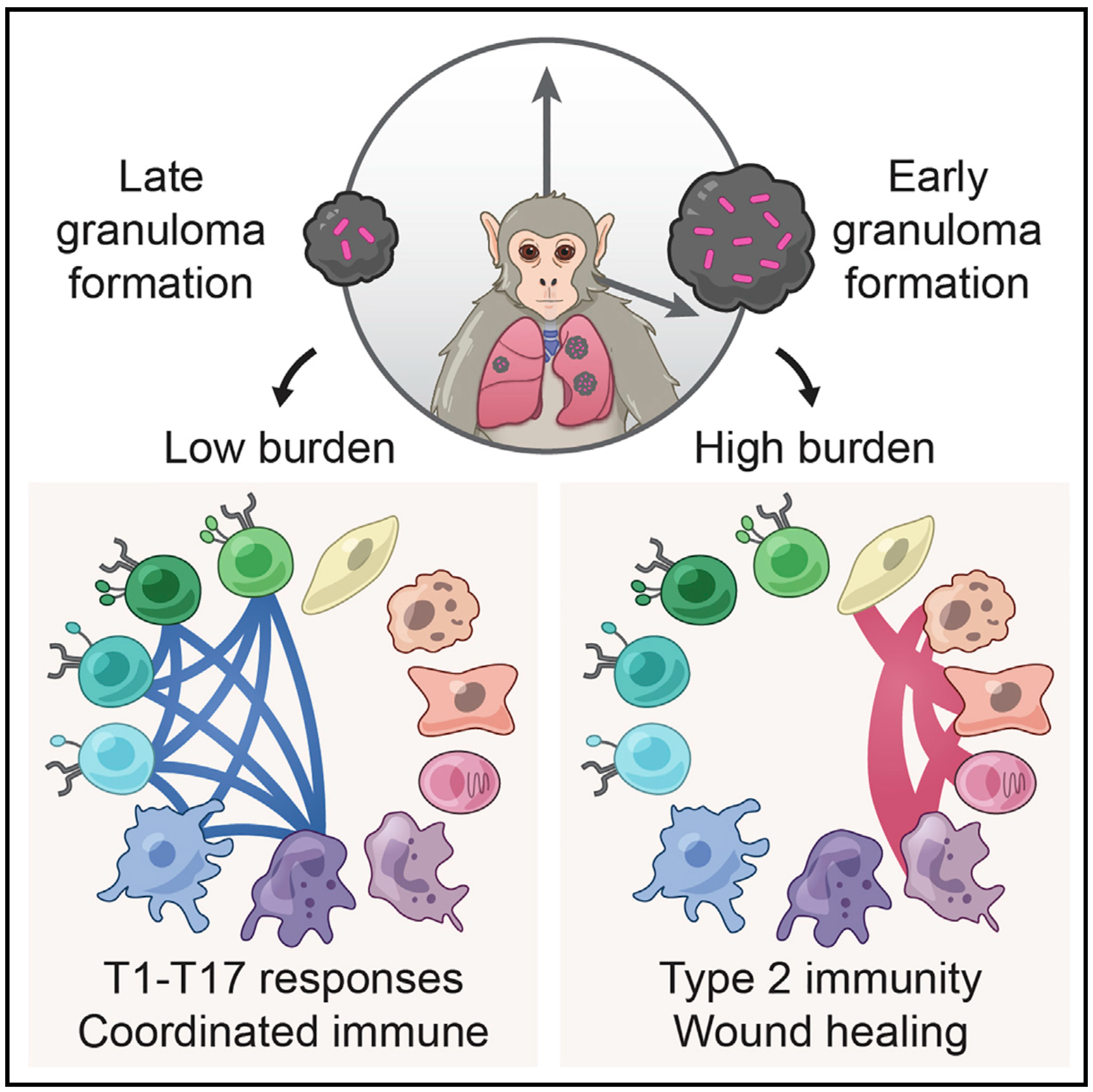
Mycobacterium tuberculosis lung infection results in a complex multicellular structure: the granuloma. In some granulomas, immune activity promotes bacterial clearance, but in others, bacteria persist and grow. We identified correlates of bacterial control in cynomolgus macaque lung granulomas by co-registering longitudinal positron emission tomography and computed tomography imaging, single-cell RNA sequencing, and measures of bacterial clearance. Bacterial persistence occurred in granulomas enriched for mast, endothelial, fibroblast, and plasma cells, signaling amongst themselves via type 2 immunity and wound-healing pathways. Granulomas that drove bacterial control were characterized by cellular ecosystems enriched for type 1-type 17, stem-like, and cytotoxic T cells engaged in pro-inflammatory signaling networks involving diverse cell populations. Granulomas that arose later in infection displayed functional characteristics of restrictive granulomas and were more capable of killing Mtb. Our results define the complex multicellular eco- systems underlying (lack of) granuloma resolution and highlight host immune targets that can be leveraged to develop new vaccine and therapeutic strategies for TB.
Malaria-causing Plasmodium vivax parasites can linger in the human liver for weeks to years and reactivate to cause recurrent blood-stage infection. Although they are an important target for malaria eradication, little is known about the molecular features of replicative and non-replicative intracellular liver-stage parasites and their host cell dependence. Here, we leverage a bioengineered human microliver platform to culture patient-derived P. vivax parasites for transcriptional profiling. Coupling enrichment strategies with bulk and single-cell analyses, we capture both parasite and host transcripts in individual hepatocytes throughout the course of infection. We define host- and state-dependent transcriptional signatures and identify unappreciated populations of replicative and non-replicative parasites that share features with sexual transmissive forms. We find that infection suppresses the transcription of key hepatocyte function genes and elicits an anti-parasite innate immune response. Our work provides a foundation for understanding host-parasite interactions and reveals insights into the biology of P. vivax dormancy and transmission.
Mycobacterium tuberculosis (Mtb) bacilli readily aggregate. We previously reported that Mtb aggregates lead to phagocyte death and subsequent efficient replication in the dead infected cells. Here, we examined the transcriptional response of human monocyte derived macrophages to phagocytosis of aggregated Mtb relative to phagocytosis of non-aggregated single or multiple bacilli. Infection with aggregated Mtb led to an early upregulation of pro-inflammatory associated genes and enhanced TNFα signaling via the NFκB pathway. These pathways were significantly more upregulated relative to infection with single or multiple non-aggregated bacilli per cell. Phagocytosis of aggregates led to a decreased phagosome acidification on a per bacillus basis and increased phagocyte cell death, which was not observed when Mtb aggregates were heat killed prior to phagocytosis. Mtb aggregates, observed in a granuloma from a patient, were found surrounding a lesion cavity. These observations suggest that TB aggregation may be a mechanism for pathogenesis. They raise the possibility that aggregated Mtb, if spread from individual to individual, could facilitate increased inflammation, Mtb growth, and macrophage cell death, potentially leading to active disease, cell necrosis, and additional cycles of transmission.
Granulomas are complex cellular structures composed predominantly of macrophages and lymphocytes that function to contain and kill invading pathogens. Here, we investigated the single-cell phenotypes associated with antimicrobial responses in human leprosy granulomas by applying single-cell and spatial sequencing to leprosy biopsy specimens. We focused on reversal reactions (RRs), a dynamic process whereby some patients with disseminated lepromatous leprosy (L-lep) transition toward self-limiting tuberculoid leprosy (T-lep), mounting effective antimicrobial responses. We identified a set of genes encoding proteins involved in antimicrobial responses that are differentially expressed in RR versus L-lep lesions and regulated by interferon-γ and interleukin-1β. By integrating the spatial coordinates of the key cell types and antimicrobial gene expression in RR and T-lep lesions, we constructed a map revealing the organized architecture of granulomas depicting compositional and functional layers by which macrophages, T cells, keratinocytes and fibroblasts can each contribute to the antimicrobial response.
Mucosal associated invariant T (MAIT) cells are a class of innate-like T cells that utilize a semi-invariant αβ T cell receptor to recognize small molecule ligands produced by bacteria and fungi. Despite growing evidence that immune cells at mucosal surfaces are often phenotypically and functionally distinct from those in the peripheral circulation, knowledge about the characteristics of MAIT cells at the lung mucosal surface, the site of exposure to respiratory pathogens, is limited. HIV infection has been shown to have a profound effect on the number and function of MAIT cells in the peripheral blood, but its effect on lung mucosal MAIT cells is unknown. We examined the phenotypic, functional, and transcriptomic features of major histocompatibility complex (MHC) class I-related (MR1)-restricted MAIT cells from the peripheral blood and bronchoalveolar compartments of otherwise healthy individuals with latent Mycobacterium tuberculosis (Mtb) infection who were either HIV uninfected or HIV infected. Peripheral blood MAIT cells consistently co-expressed typical MAIT cell surface markers CD161 and CD26 in HIV-negative individuals, while paired bronchoalveolar MAIT cells displayed heterogenous expression of these markers. Bronchoalveolar MAIT cells produced lower levels of pro-inflammatory cytokine IFN-γ and expressed higher levels of co-inhibitory markers PD-1 and TIM-3 than peripheral MAIT cells. HIV infection resulted in decreased frequencies and pro-inflammatory function of peripheral blood MAIT cells, while in the bronchoalveolar compartment MAIT cell frequency was decreased but phenotype and function were not significantly altered. Single-cell transcriptomic analysis demonstrated greater heterogeneity among bronchoalveolar compared to peripheral blood MAIT cells and suggested a distinct subset in the bronchoalveolar compartment. The transcriptional features of this bronchoalveolar subset were associated with MAIT cell tissue repair functions. In summary, we found previously undescribed phenotypic and transcriptional heterogeneity of bronchoalveolar MAIT cells in HIV-negative people. In HIV infection, we found numeric depletion of MAIT cells in both anatomical compartments but preservation of the novel phenotypic and transcriptional features of bronchoalveolar MAIT cells.
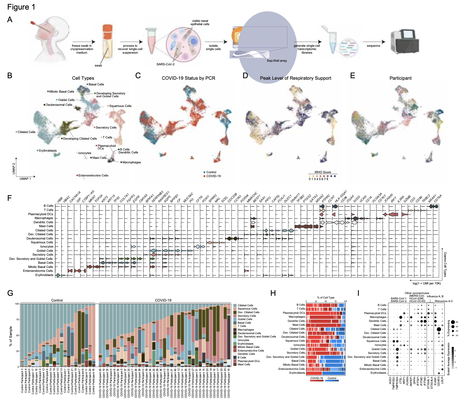
Infection with SARS-CoV-2, the virus that causes COVID-19, can lead to severe lower respiratory illness including pneumonia and acute respiratory distress syndrome, which can result in profound morbidity and mortality. However, many infected individuals are either asymptomatic or have isolated upper respiratory symptoms, which suggests that the upper airways represent the initial site of viral infection, and that some individuals are able to largely constrain viral pathology to the nasal and oropharyngeal tissues. Which cell types in the human nasopharynx are the primary targets of SARS-CoV-2 infection, and how infection influences the cellular organization of the respiratory epithelium remains incompletely understood. Here, we present nasopharyngeal samples from a cohort of 35 individuals with COVID-19, representing a wide spectrum of disease states from ambulatory to critically ill, as well as 23 healthy and intubated patients without COVID-19. Using standard nasopharyngeal swabs, we collected viable cells and performed single-cell RNA-sequencing (scRNA-seq), simultaneously profiling both host and viral RNA. We find that following infection with SARS-CoV-2, the upper respiratory epithelium undergoes massive reorganization: secretory cells diversify and expand, and mature epithelial cells are preferentially lost. Further, we observe evidence for deuterosomal cell and immature ciliated cell expansion, potentially representing active repopulation of lost ciliated cells through coupled secretory cell differentiation. Epithelial cells from participants with mild/moderate COVID-19 show extensive induction of genes associated with anti-viral and type I interferon responses. In contrast, cells from participants with severe lower respiratory symptoms appear globally muted in their anti-viral capacity, despite substantially higher local inflammatory myeloid populations and equivalent nasal viral loads. This suggests an essential role for intrinsic, local epithelial immunity in curbing and constraining viral-induced pathology. Using a custom computational pipeline, we characterized cell-associated SARS-CoV-2 RNA and identified rare cells with RNA intermediates strongly suggestive of active replication. Both within and across individuals, we find remarkable diversity and heterogeneity among SARS-CoV-2 RNA+ host cells, including developing/immature and interferon-responsive ciliated cells, KRT13+ “hillock”-like cells, and unique subsets of secretory, goblet, and squamous cells. Finally, SARS-CoV-2 RNA+ cells, as compared to uninfected bystanders, are enriched for genes involved in susceptibility (e.g., CTSL, TMPRSS2) or response (e.g., MX1, IFITM3, EIF2AK2) to infection. Together, this work defines both protective and detrimental host responses to SARS-CoV-2, determines the direct viral targets of infection, and suggests that failed anti-viral epithelial immunity in the nasal mucosa may underlie the progression to severe COVID-19.
Influenza virus infections are major causes of morbidity and mortality. Research using cultured cells, bulk tissue, and animal models cannot fully capture human disease dynamics. Many aspects of virus-host interactions in a natural setting remain unclear, including the specific cell types that are infected and how they and neighboring bystander cells contribute to the overall antiviral response. To address these questions, we performed single-cell RNA sequencing (scRNA-Seq) on cells from freshly collected nasal washes from healthy human donors and donors diagnosed with acute influenza during the 2017-18 season. We describe a previously uncharacterized goblet cell population, specific to infected individuals, with high expression of MHC class II genes. Furthermore, leveraging scRNA-Seq reads, we obtained deep viral genome coverage and developed a model to rigorously identify infected cells that detected influenza infection in all epithelial cell types and even some immune cells. Our data revealed that each donor was infected by a unique influenza variant and that each variant was separated by at least one unique non-synonymous difference. Our results demonstrate the power of massively-parallel scRNA-Seq to study viral variation, as well as host and viral transcriptional activity during human infection.
Toxoplasma gondii chronically infects a quarter of the world’s population, and its recrudescence can cause life-threatening disease in immunocompromised individuals and recurrent ocular lesions in the immunocompetent. Acute-stage tachyzoites differentiate into chronic-stage bradyzoites, which form intracellular cysts resistant to immune clearance and existing therapies. The molecular basis of this differentiation is unknown, despite being efficiently triggered by stresses in culture. Through Cas9-mediated screening and single-cell profiling, we identify a Myb-like transcription factor (BFD1) necessary for differentiation in cell culture and in mice. BFD1 accumulates during stress and its synthetic expression is sufficient to drive differentiation. Consistent with its function as a transcription factor, BFD1 binds the promoters of many stage-specific genes and represents a counterpoint to the ApiAP2 factors that dominate our current view of parasite gene regulation. BFD1 provides a genetic switch to study and control Toxoplasma differentiation and will inform prevention and treatment of chronic infections.
Toxoplasma gondii chronically infects a quarter of the world’s population, and its recrudescence can cause life-threatening disease in immunocompromised individuals and recurrent ocular lesions in the immunocompetent. Chronic stages are established by differentiation of rapidly replicating tachyzoites into slow-growing bradyzoites, which form intracellular cysts resistant to immune clearance and existing therapies. Despite its central role in infection, the molecular basis of chronic differentiation is not understood. Through Cas9-mediated genetic screening and single-cell transcriptional profiling, we identify and characterize a putative transcription factor (BFD1) as necessary and sufficient for differentiation. Translation of BFD1 appears to be stress regulated, and its constitutive expression elicits differentiation in the absence of stress. As a Myb-like factor, BFD1 provides a counterpoint to the ApiAP2 factors which dominate our current view of parasite gene regulation. Overall, BFD1 provides a genetic switch to study and control Toxoplasmadifferentiation, and will inform prevention and treatment of chronic infection.
As many factors define cellular phenotype and influence disease beyond mRNA, we develop complementary methods for co-assaying other cellular attributes to enrich our understanding of the drivers of cellular behaviors. Examples including the abundance of additional ‘-omes’, the sequence and amount of important transcripts, cellular history, biophysical properties, spatial position, and functional output. Recently, we have worked to: 1. detect pathogens in cells and potentially actionable associated host factors; 2. query for specific mutations to identify cancer cells; and, 3. extract T cell receptor sequences to examine clonality. We have also formulated computational methods to derive deeper insights from these data (e.g., to examine viral dynamic in infected cells, reproducible features hidden by inter-individual variability, multicellular immune dynamics, intercellular communication, or alteration in cellular ecosystems associated with pathology).
We explore how the extracellular milieu influences cellular decision-making. Here, we have employed controlled culture conditions with cells and organoids, chemical and genetic perturbations, and constant microfluidic perfusion. We also have leveraged natural microenvironmental variation within and across tissues via microdissection and by using photoactivatable probes that retain spatial information through dissociation. In each instance, we aim to understand the degree to which extracellular environments modulate, and can be used to rationally control, the responses of individual cells or the overall distribution thereof, with an eye toward engineering tissue responses.
We examine the impact of intercellular interactions on cellular function. We have used coculture, imaging and perturbation strategies, as well as matched computational methods, to reinforce findings from dissociated samples, validate inferred cell-cell communication in vivo (e.g., between sensory neurons and lymph node resident cells), and manipulate multicellular systems (e.g., organoids). We are currently working on building arrayed, synthetically-designed cellular ensembles to examine how ‘tissue’ structure impacts functional response. Our overall goal is to understand cellular co-dependencies that influence niche- and tissue-level response dynamics.
Vaccines remain the most effective tool to prevent infectious diseases. Here, we introduce an in vitro booster vaccination approach that relies on antigen-dependent activation of human memory B cells in culture. This stimulation induces antigen-specific B cell proliferation, differentiation of B cells into plasma cells, and robust antibody secretion after a few days of culture. We validated this strategy using cells from healthy donors to retrieve human antibodies against tetanus toxoid and influenza hemagglutinin (HA) from H1N1 and newly emergent subtypes such as H5N1 and H7N9. Anti-HA antibodies were cross-reactive against multiple subtypes, and some showed neutralizing activity. Although these antibodies may have arisen as a result of previous influenza infection, we also obtained gp120-reactive antibodies from non–HIV-infected donors, indicating that we can generate antibodies without prior antigenic exposure. Overall, our novel approach can be used to rapidly produce therapeutic antibodies and has the potential to assess the immunogenicity of candidate antigens, which could be exploited in future vaccine development.
We lack effective treatments and preventions for many of the most challenging infectious diseases, many of which disproportionately impact those in low- and middle-income countries or traditionally marginalized communities.
To help address this, we have established and enabled multi-group, multi-country partnerships to deploy and adapt cutting-edge genomic tools. By examining how cells dynamically alter their states, individually and collectively, during disease and/or its resolution in acute and chronic infections—e.g., tuberculosis, HIV/SHIV, hepatitis, malaria, leprosy, flu, SARS-CoV-2, and ebola—we have uncovered cellular and molecular features of pathogen control or pathology to potentiate or counteract, respectively. Illustratively, in tuberculosis, we identified a functional role for cytotoxic CD8 and hybrid type1-type17 T cells in control of infection in the lung and links between mast, plasma, and endothelial cell abundance (type-2 immune responses) and bacterial burden. We have also built methods for examining pathogens within individual host cells to define their dynamic interdependence and identify potentially restrictive host factors.
We are currently working to identify the drivers of common host responses to distinct perturbations and their targetability, as well as the impact of different interventions (e.g., vaccines).
Immune responses play a critical role in preventing tumorigenesis. Sometimes, however, they are ineffectual and can even drive/support malignancy.
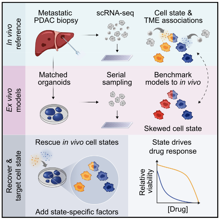
We have examined how cancer cells alter and are influenced by their tumor microenvironments (TMEs), and the impact this has on therapeutic responses. Illustratively, in Pancreatic Ductal Adenocarcinoma (PDAC), by profiling liver metastases and matched organoid models, we showed: 1. associations between TME and malignant cell state composition; 2. that autocrine and paracrine signaling can drive malignant cell state transitions, even in an isogenic background, altering the efficacy of frontline chemotherapies; and, 3. that microenvironmental manipulations can be used to control malignant state, and thereby drug responses, rationally, and to improve model fidelity for screening potential therapies. This and related work highlight the potential utility of modulating indirect target cells (T cells in the PDAC TME or basal cells in allergic inflammation) to enhance cures and preventions.
We are now systematically expanding this work to define how additional environmental and cell-intrinsic factors influence malignant cell state plasticity in PDAC and other cancers toward enhancing treatments.
Encounters between immune cells and invading bacteria ultimately determine the course of infection. These interactions are usually measured in populations of cells, masking cell-to-cell variation that may be important for infection outcome. To characterize the gene expression variation that underlies distinct infection outcomes and monitor infection phenotypes, we developed an experimental system that combines single-cell RNA-seq with fluorescent markers. Probing the responses of individual macrophages to invading Salmonella, we find that variation between individual infected host cells is determined by the heterogeneous activity of bacterial factors in individual infecting bacteria. We illustrate how variable PhoPQ activity in the population of invading bacteria drives variable host type I IFN responses by modifying LPS in a subset of bacteria. This work demonstrates a causative link between host and bacterial variability, with cell-to-cell variation between different bacteria being sufficient to drive radically different host immune responses. This co-variation has implications for host-pathogen dynamics in vivo.